Large animal - October 2018
Bovine caesareans: alternative approaches to difficult cases
Emmet Kelly MVB and European College Resident in Ruminant Reproduction, University College Dublin, and Eoin Ryan MVB MVM, diplomate of the European College of Bovine Health Management and assistant professor in Farm Animal Clinical Studies, University College Dublin, outline alternative approaches to bovine caesarean
Introduction
With the commencement of the autumn calving season, caesarean sections will again become a common occurrence for the management of dystocia both in mixed- and farm-animal practice. As most practitioners are comfortable in managing routine caesareans, the following article aims to outline some alternative approaches to dealing with more challenging caesarean sections such as those on fractious cows or those with extremely heavy and/or emphysematous calves.
Case selection
As most practitioners are aware, caesareans have multiple indications and complications, and these are outlined in Table 1. Depending on the indication for the caesarean, the complications and outcome for the cow, calf and farmer can differ considerably.
|
Indications for surgery |
Common complications |
|
Elective for high-value animals |
Peritonitis |
|
Foetomaternal disproportion (relative/absolute) |
Subcutaneous emphysema |
|
Incomplete dilation of the cervix/vagina/vulva |
Adhesions |
|
Irreducible uterine torsion |
Wound dehiscence, abscesses or seromas |
|
Inability to correct foetal maldisposition |
Postpartum haemorrhage |
|
Inability to perform foetotomy on an emphysematous calf |
Retained foetal membranes |
|
Inability to perform foetotomy on foetal monsters |
Infertility |
Table 1: Common indications for and complications of caesareans.
The viability of the cow and calf at the time of surgery is widely accepted as a major determining factor in the overall outcome. For that reason, in a review of bovine caesareans in 2005, Newman and Anderson, proposed to differentiate caesareans into three types: elective, emergency with a non-emphysematous foetus; and emergency with an emphysematous foetus, as the anticipated outcomes and complications dramatically differ between each situation.
For example, once an emphysematous foetus is present, the dam survival rate drops to 33% compared to 86-98% survival rates quoted for uncomplicated caesareans with a live calf. This is likely due to the fact these cows are toxic, pyrexic, hypotensive and that the risk of abdominal contamination during surgery is high. Of course, there are a multitude of other factors that also influence the outcome and most of the major ones are outlined below in Table 2.
|
Cow- and calf-based risk factors |
Common complications |
|
Health of cow at initiation of surgery |
Elective or emergency |
|
Presence of a live, dead or emphysematous foetus |
Ability to exteriorise the uterus ( dam survival) |
|
Sex of the foetus (female: cow survival) |
Surgical time (>1 hour: dam survival) |
|
Age/parity (primiparous: survival) |
Surgeon skill/experience |
|
Type of cow (beef: dam and calf survival) |
Hygiene of surgeon and surgical environment |
|
Temperament of the dam |
Degree of perioperative obstetric manipulation/rapid decisionmaking ( dam survival) |
|
Duration of dystocia |
Removal of abdominal blood clots ( dam survival |
| Type of dystocia (foeto-maternal disproportion: cow survival) | Use of Utrecht suture pattern ( culling risk) |
| Condition of the uterus (oedematous or friable) | Availability of skilled help |
| Retained foetal membranes following surgery | Use of paravertebral anaesthesia ( culling risk) |
Table 2: Main factors affecting overall outcomes for caesareans.
Irrespective of the nature of the dystocia case that you are presented with, it is important to choose cases for caesareans carefully with your safety, the welfare of the cow and the economics of the situation always to the forefront of your mind.
Handling facilities
The handling facilities that are available on-farm will have a huge bearing on the way caesareans are approached and in their eventual outcome. In an ideal world, all cows should be restrained in a crush or a calving gate like those in Figure 1. However, the reality is that some crushes prevent surgical access and/or have no way of releasing cows that become recumbent.
If the cow is agreeable, many can be managed standing with assistance holding the tail upright if they are loose haltered to a fixed point in a shed. However, even in quiet cows, swaying over and back can be a nuisance and unsafe. Sedation (as outlined below) can be useful in these cases. If possible, single or double-haltering cows halfway up a crush or a race and placing a fixed bar in front of them to stop them surging forward can restrain, stop side-to-side movement and give good access to the surgical site. Sometimes, placing a loose rope on the right hind limb in cows that have the potential of going down can facilitate pulling this limb from under the cow to keep the incision site off the ground. From a personal safety point of view, avoid doing caesareans where the gates or posts that are being used to restrain are poorly fixed; where there is risk of getting pinned between a wall/gate; and where a quick escape is not possible if the cow breaks loose from her restraints. Recumbent sedation with rope restraint is often the only safe method of operating on wild cows where facilities are lacking. Nevertheless, always be careful as sedation can be unreliable and ropes can break so ensure when possible you are not in a kick-zone and can escape easily.
Sedative options
The key things to consider when choosing the appropriate sedation protocol are the health and temperament of the animal; the time required for the procedure; whether or not recumbency is required; and the handling facilities and help on farm. All of these factors will dictate what method of sedation is appropriate. The options for both standing and recumbent sedation for surgery are outlined in Tables 3 and 4, respectively. In Ireland, the anaesthetic drugs that are licenced for use in cattle are limited, thus restricting practitioners’ options. It is worth noting that butorphanol is listed on both Tables 3 and 4 and although it is not licensed in cattle, it does have a licence for food-producing equines and can be used on the cascade in cattle once a 28-day meat withdrawal and seven-day milk withdrawal is observed.
| Options for standing sedation |
Dose range (mg/kg) |
Standard dose range (ml/100kg) |
Duration of effect | Fractious cattle dose (ml/100kg) | Comments |
| Xylazine 2% only | 0.015-0.1mg/kg (IM) 0.0075-0.05mg/kg (IV) |
0.075-0.2ml (IM) 0.0375-0.1ml (IV) |
30-45 mins |
0.2-0.5ml (IM) 0.1-0.25ml (IV) |
IV dose is half IM dose |
|
Detomidine 1% only |
0.006-0.02mg/kg (IM) 0.002-0.015mg/kg (IV) |
0.06-0.1ml (IM) 0.02-0.05ml (IV) |
30-60 mins |
0.15-0.2ml (IM) 0.1-0.15ml (IV) |
Cardiorespiratory depressant – not for recumbency |
| Xylazine-lidocaine epidural | 0.015-0.07mg/kg (Xylazine) |
0.25-0.5ml per 300kg | 45-60 mins | 0.5-1ml per 300kg | Make up to 5ml with local anaesthetic |
| Ketamine stun options | Dose range (mg/kg) | Standard dose range (ml/100kg) |
Duration of effect |
Fractious cattle dose (ml/100kg) |
Comments |
| IV standing stun Butorphanol Xylazine Ketamine |
0.02-.1mg/kg 0.02-0.0275mg/kg 0.05-0.1mg/kg |
0.2-1ml 0.1-0.15ml 0.05-0.1ml |
15 mins | As per standard dose range | Suited to short procedures |
| SQ/IM standing stun 5-10-20 Butorphanol Xylazine Ketamine |
0.01mg/kg(IM/SQ) 0.02mg/kg(IM/SQ) 0.04mg/kg(IM/SQ) |
0.1ml 0.1ml 0.04ml |
60-90 mins |
10-20-40 Stun 0.2ml 0.2ml 0.08ml |
Get longer duration & less recumbency with SQ route Can ketamine to 0.2ml/100kg if very wild & give IM but beware of recumbency at high doses |
|
Stun without butorphanol Xylazine Ketamine |
0.04mg/kg(IM/SQ) 0.2mg/kg(IM/SQ) |
0.2ml 0.2ml |
60-90 mins |
0.4-0.6ml 0.4-0.6ml |
Not as reliable as standard stun in wild cattle |
Table 3: Sedative options for standing surgery in the cow.
|
Options for recumbent sedation |
Dose range (mg/kg) |
Standard cattle dose (ml/100kg) |
Duration of effect |
Fractious cattle dose (ml/100kg) |
Comments |
| Xylazine 2% only |
0.2-0.4mg/kg (IM) 0.1-0.2mg/kg (IV) |
1ml (IM) 0.5ml (IV) |
30-45mins <30mins |
1.5-2ml (IM) 0.75-1ml (IV) |
May still need to cast to induce recumbency |
|
Xylazine 2% + Ketamine |
0.05-0.1mg/kg (IM) 0.025-0.05mg/kg (IV) + 4mg/kg (IM) 2mg/kg (IV) |
0.25ml (IM) 0.125ml (IV) + 4ml (IM) 2ml (IV) |
30-40 mins (IM) 5-10 mins (IV) |
0.5ml (IM) 0.25ml (IV) + 4ml (IM) 2ml (IV) |
IM can be given together IV option Xylazine given 5 minutes before Often need to top up ketamine in half doses |
|
IV recumbent Stun Butorphanol Xylazine Ketamine |
0.05-0.1mg/kg 0.025-0.05mg/kg 0.3-0.5mg/kg |
0.5ml 0.125ml 0.3ml |
15 mins |
1ml 0.25ml 0.5ml |
Only suited to very short procedures - <25mins even with top-ups |
|
SQ/IM recumbent stun Butorphanol Xylazine Ketamine |
0.025mg/kg 0.05mg/kg 0.1mg/kg |
0.25ml 0.25ml 0.1ml |
30-45 mins |
0.5ml 0.5ml 0.2ml |
Longer when given SQ From a practical point of view, if facilities are good and/or the dam is agreeable, recumbent or standing sedation is not required. However, if the dam is wild, strong-standing sedation or a recumbent approach with sedation can be a safer alternative. Also, if the crush is absent/inappropriate; if the cow has the potential to go down or if the calf is very large/emphysematous, inducing recumbency should be strongly considered. It is also worth noting that it is much better to start a caesarean in a recumbent position rather than starting in a standing position followed by the cow becoming recumbent mid-surgery, as these cows are at a much higher risk of peritonitis, wound infections or death. |
Table 4: Sedative options for recumbent surgery in the cow
For both caesarean sections and foetotomies, it can be useful if the cow stops straining and is mildly sedated. The xylazine-lidocaine epidural can work very well in this respect and the lower dose (0.5ml per 300kg) could be considered even in routine caesareans to reduce straining, switching of the tail and to facilitate manipulations during surgery. Clenbuterol (10ml) can be given intravenously (IV) to facilitate manipulation of the calf during surgery and it also counteracts the ecbolic effects of alpha-2-adrenergic agonists (xylazine/detomidine) used for sedation.
The intramuscular or subcutaneous standing ketamine ‘stun’ is the preferred method of standing sedation for caesarean section. The standard stun or the 5-10-20 stun (so-called because 5mg of butorphanol, 10mg of xylazine and 20mg of ketamine are the amounts in mgs given to a 500kg cow) given together subcutaneously is appropriate in moderately anxious cows. Supplemental amounts of 25% to 50% of the initial xylazine and ketamine doses can be used as top ups if further time or co-operation is needed. In very fractious cows the intramuscular route is preferred and the 10-20-40 stun works well. For practitioners that are reticent about using butorphanol, the modified ketamine stun, ie. 1ml xylazine 2% together with 1ml ketamine, works well in most cows. Only in extremely wild cows would a surgeon need to administer a dose as high as 2ml xylazine 2% and 2ml ketamine and the risk of recumbency increases at this dose.
The reason for inducing recumbency is very important in terms of what drug/s and doses are chosen. If it is decided to do the caesarean in a recumbent position in a normal healthy cow, due to a lack of facilities for example, xylazine alone or xylazine-ketamine intramuscular options are adequate. However, be aware that the use of high doses of sedatives to achieve recumbency can impair the viability of the calf and the surgeon must work quickly to block the cow and retrieve the calf. If the cow is in a compromised state (toxaemic, hypotensive or in shock), such as in the case of those cows carrying emphysematous calves, then using low doses of all drugs (especially xylazine 2%) is strongly recommended. The standard subcutaneous recumbent stun is probably the least likely to cause cardiorespiratory depression. Rope restraint of all legs is essential and the ropes should be tied to a fixed point with a quick-release knot. Regardless of the sedative options chosen, local anaesthetic blocks (paravertebral, inverted-L or line) should be still conducted prior to surgery.
Surgical approaches for difficult cases
The left paralumbar celiotomy is by far the most recognised, standardised and appropriate approach used in standing uncomplicated caesarean sections and will not be discussed any further in this article.
Standing right paralumbar celiotomy
Most practitioners are familiar with right sided approaches from displaced abomasal surgery; however, in terms of the incision, it is much larger and made in a similar location to the left sided celiotomy (mid-paralumbar fossa 10cm ventral to the transverse processes). Due to the difficulty of keeping the intestines in the peritoneal cavity, this approach is not recommended routinely. This approach is advantageous, however, if there is a lot of scar tissue/adhesions on the left side in a cow that has had repeated caesareans, or if a very large calf is present in the right horn and there is genuine concern about manipulating the calf from right to left.
Recumbent ventral celiotomy approaches (midline and paramedian)
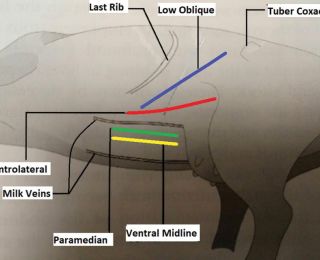
The two ventral approaches are the ventral midline and paramedian approaches. The cow is sedated and put in dorsal recumbency leaning 45° towards the surgeon with both feet tied. The midline incision is made through the skin, fascia and linea alba starting 5-7cm cranial to umbilicus and extending as caudal as required, while the paramedian is made 5cm lateral to linea alba and medial to the milk vein (Figure 2). The uterus is exteriorised by untying the hind limbs and temporally laying them flat on the ground (so the cow is in lateral recumbency) and once the foetus is removed the legs are tied back in place. The inner layer of the incision is closed incorporating the linea alba or rectus abdominis sheath as appropriate, and the musculature with vicryl in a simple continuous or a horizontal mattress pattern. Although both methods offer good access and exteriorisation of the uterus and could be used for emphysematous calf removal, they are problematic for a number of reasons: heavy sedation and manpower are required to put and keep the cow in dorsal recumbency; the abdominal incision can be difficulty to close; and there is a considerable risk of abdominal wall herniation or evisceration if abdominal wall closure is poor. Wound infection is more of an issue due to the ventral location also.
Recumbent ventrolateral celiotomy (Hannover approach)
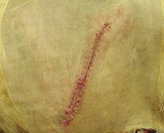
The cow is placed in right lateral recumbency (Figure 2) and the legs are tied, with the left leg extended caudally and abducted to get good exposure to the incision and the uterus. A curved incision is made starting 20cm dorsal to the attachment of udder, medial to the fold of the stifle, and it is continued about 50cm cranioventrally to allow exteriorisation of the gravid horn (Figure 2). Once exteriorised, the gravid horn is incised along its ventral aspect, the calf is removed, and the uterus sutured using absorbable suture material in one or two layers (modified Cushing also known as Utrecht method) depending on whether it is an emphysematous foetus or not. The muscles are closed in three layers with six to eight metric absorbable-suture material in a continuous pattern. The advantages of this method are that it avoids the well vascularised musculature of the flank, does not require dorsal recumbency and allows excellent exteriorisation of the uterus. Its disadvantages are that the closure can be difficult due to tension, wound infection is more likely due to location and the integrity of the abdominal closure is less secure than that of either the ventral midline or paramedian approaches.
Standing/recumbent left oblique celiotomy (Liverpool approach)
The incision starts 4-6cm ventral and cranial to the tuber coxa and is extended cranioventrally at a 45°-angle toward the caudal rib where it stops (Figure 3). As it goes more cranial and ventral than the classic left paralumbar approach, the apex of the gravid horn is more readily exteriorised. The external muscle layer is incised in the same direction of the skin while the internal abdominal oblique and transverse abdominal muscles can then be gridded parallel to the incision with sharp and blunt dissection. The uterus is incised usually along the greater curvature and closed as per normal in one or two layers as above. The muscles are closed in a three-layer closure as described above. This approach allows easier access to the uterus and can be particularly useful if there is a very heavy calf present as it requires less physical strength to exteriorise the calf. It does not require much more help than what is required of for a standard standing-left approach and there is much less risk of herniation or evisceration. This method can also be used in a recumbent approach with the incision starting lower, about 8-10cm ventral and cranial to the tuber coxa, and if the cow is rolled into near sternal it will facilitate exteriorisation of the uterus. The recumbent approach is more appropriate than the standing method for the emphysematous foetus as it results in less abdominal contamination and better exteriorisation of the uterus.
The emphysematous case
Foetotomy is the preferred method to remove emphysematous calves but it is not always possible. As previously mentioned, deciding whether to conduct a caesarean or to euthanise the cow is not a simple one and will depend on many factors. Caesarean section is, undoubtedly, a salvage operation as the probability of a subsequent successful pregnancy in those cows that survive is estimated to be less than 25%. In 2013 in the UK, Dale et al. described cases that they had operated on in the field and estimated that, for the surgery to be economically feasible in dairy cows, survival rates had to be 50% or greater. Of the six cases they described, four lived (66% survival rate), while Bouchard (1994) only had a 33% survival rate out of 16 cases. This highlights that, for these caesareans to make economic sense, both case selection and optimal management of the cow are vital.
Preoperative management of the cow is paramount. The foetus is not going anywhere at this stage, so often it may be better to stabilise the cow before rushing into surgery. This involves aggressive fluid therapy with oral fluids (30-50L), antibiotics and non-steroidal anti-inflammatory drugs (NSAIDs) if the cow is systemically sick or endotoxaemic. IV fluid therapy should also be considered if the cow is in shock as fluid absorption from the rumen of hypovolemic cows can be poor. Large volumes (40-60L) of isotonic intravenous fluid given over 12-24 hours would help but is likely to be impractical and costly. Alternatively, a hypertonic saline (7.2%) drip given at a rate of 5ml/kg over a four-minute period (which is approximately 3L for an average adult cow) is more practical but it must be combined with oral fluid administration. The uterine environment of these cows is heavily contaminated by a polymicrobial population consisting of Truepurella pyogenes, Escherichia coli, Fusobacterium spp, Proteus spp and Bacteroides spp and, therefore, broad spectrum antibiotics, such as oxytetracycline (20mg/kg IV), and NSAIDs, eg. flunixin (1mg/kg IV), are advised pre-operatively.
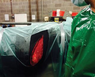
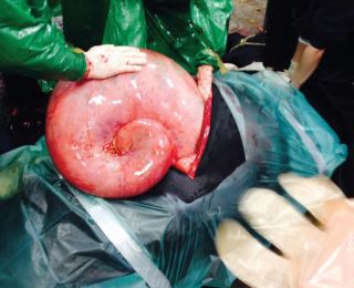
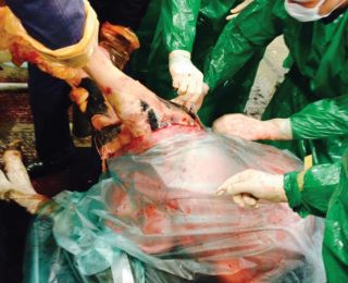
Once the cow is stabilised, surgery may be undertaken. Surgery to remove an emphysematous foetus should be considered a two-vet operation in all cases. The standing left oblique and any of the ventral approaches (midline, paramedian, ventrolateral or the low oblique flank) are appropriate but, recumbent ventrolateral or recumbent low oblique are preferred in a farm setting. Whatever method used, good exteriorisation of the uterus is critical to prevent contamination of the abdomen (Figure 4). As the cow is recumbent, draping with a sterile disposable drape is advised and, once the uterus is exteriorised, it should also be draped, and the uterine incision made through this to reduce contamination (Figure 4). The placental membranes should be removed if they are easily detached from the maternal caruncles (which is common due to their lytic state) and the incision sutured with a two-layer closure using absorbable suture material in the modified Cushing (Utrecht) suture pattern. The uterine surface should then be thoroughly lavaged with saline (Figure 5). At this point, re-scrubbing and changing gloves and kits is advised to try and reduce contamination of the surgical site. The uterus is then placed back in the abdomen and the muscle layers and skin closed in the appropriate way depending on the approach taken.
Post-operatively, the cow should receive further oral and possibly IV fluids and be maintained on antibiotics for a minimum of a week and NSAIDs for three days. The cow should be housed separately in a clean pen for four weeks.
Key points
- The overall outcome of a caesarean is influenced by a range of factors:
- Good handling facilities and help on farm are key to ensuring safety;
- Sedation can be used to facilitate handling of anxious/unruly cows and to induce recumbency;
- Left paralumbar celiotomy remains the recommended approach for most caesareans;
- A caesarean on an emphysematous case is a salvage procedure and should only be considered in stabilised cows where it is deemed there is a reasonable chance of success;
- Many approaches are available to deal with the emphysematous case each with its advantages and disadvantages; and
- Good exteriorisation of the uterus, minimising abdominal contamination and diligent supportive care are the key to achieving better outcomes for these cases.
- Abrahamsen EJ (2008) Ruminant Field Anaesthesia. Vet Clin North Am Food Anim Pract 24: 429-441
- Alexander D (2013) Bovine caesarean section 1. On-farm operations. In Practice 35: 574-588
- Alexander D (2013) Bovine caesarean section 2. Difficult caesareans (potential pitfalls and how to overcome them). In Practice 36: 15-26
- Bouchard E, Daignault D, Bélanger D, Couture Y (1994) Caesareans on dairy Cows: 159 cases. Can Vet J 35(12): 770-4
- Dale H, Hayes E, DeKruif A (2013) Case study: the use of a salvage (low flank) caesarean section to remove an emphysematous calf. Livestock 18(5): 175-178
- Funnell BJ, Hilton WM (2016) Management and Prevention of Dystocia Vet Clin North Am Food Anim Pract 32: 511-522
- Noakes DE, Parkinson TJ, England GCW (2009) Veterinary Reproduction an Obstetrics, Ch. 20, 9th Edition. Elsevier Ltd.: 347-366
- Newman KD, Anderson DE (2005) Caesarean Section in Cows. Vet Clin North Am Food Anim Pract 21: 73-100
- Schultz LG, Tyler JW, Moll HD, Constantinescu GM (2008) Surgical Approaches for Caesarean Section in Cattle. Can Vet J 49(6): 565-8


