Overview of vertebral fractures in small animals
In this article, Ronan A Mullins MVB DECVS PGDipUTL DVMS MRCVS, EBVS European specialist in small animal surgery, associate professor and discipline head of small animal surgery at University College Dublin, gives an overview of vertebral fractures in small animals and an insight into research he is undertaking into ways of improving the safety of spinal instrumentation
Vertebral fractures are an important cause of spinal cord injury in dogs. The majority are due to severe blunt trauma, most commonly secondary to road traffic accidents (~ 40-60 per cent of cases). Other possible causes include falls, kicks, and animal attacks, and less commonly secondary to underlying pathology such as vertebral neoplasia, infection or metabolic bone disease. Fractures can affect any region of the vertebral column but the most common regions are T3-L3 and L4-S3. Vertebral fractures can result in primary and secondary spinal cord injury. Primary spinal cord injury relates to the mechanical insult, whereas secondary spinal cord injury is related to the cascade of events that follows involving free radicals, inflammatory mediators, cytokines, excitatory neurotransmitters, and so forth.
It is important to remember when presented with an animal with a suspected vertebral fracture not to overlook the possibility of concurrent life threatening injuries. On the basis of the traumatic nature of vertebral fractures, it is not surprising that ~ 45-85 per cent have concurrent injuries. These can include thoracic injuries such as pneumothorax, pulmonary contusions, fractured ribs; abdominal injuries such as bladder rupture, haemoabdomen, organ injury; pelvic and long bone fractures; and so forth. Therefore, a thorough assessment of the patient should be performed, including thoracic and abdominal assessment with sonography for trauma (T-FAST and A-FAST, respectively), and a minimum database should be obtained, consisting of haematology, biochemistry and blood gas.
Patients presenting with signs of hypovolaemic shock should be provided with intravenous fluid therapy, correcting for any electrolyte abnormalities. Appropriate analgesia should be provided but ideally after neurologic assessment has been performed to avoid interference therewith and making interpretation of findings difficult. Patients presenting with vertebral injury and at risk of having suffered a vertebral fracture should be immobilised to prevent further injury to the spinal cord. In some cases, light sedation may be required if immobilisation is poorly tolerated and resulting in thrashing behaviour. A thorough neurologic examination should be performed to determine the neurolocalisation and the severity of spinal cord injury.
Nociception
The presence of nociception is the single most important prognostic factor for neurologic recovery following spinal cord injury. Absence of nociception in the limbs caudal to the spinal cord lesion (e.g., absence of nociception in the pelvic limbs with a T3-L3 spinal cord injury) indicates a very poor prognosis for return of function. It is important to remember that the patient must demonstrate a conscious response to the noxious stimulus (e.g., forceps pinching the toes or tail) such as a yelp, turning of the head toward the stimulus, or wanting to bite. Simply withdrawing the limb does not indicate the presence of nociception. In comparison to dogs with grade 5 spinal cord injury (para/tetra-plegia with absence of nociception) secondary to intervertebral disc herniation who have a ~ 50-60 per cent rate of neurologic recovery with surgery, those with this severity of spinal cord injury secondary to vertebral fracture have a very grave prognosis. In the majority of such cases, euthanasia is recommended because of expected permanent paralysis, lifelong bladder and bowel incontinence, pressure sores, and recurrent urinary tract infections secondary to incomplete bladder emptying and recumbency.
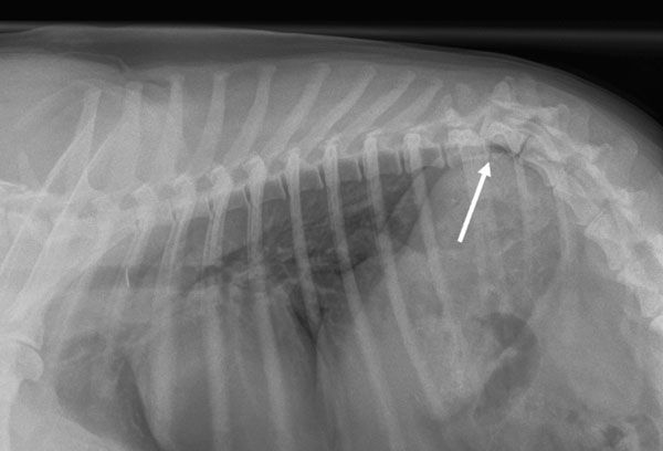
Figure 1: Lateral thoracic radiograph demonstrating a significantly displaced T11-12 vertebral fracture in a Collie following a road traffic accident. On neurologic examination, the dog was paraplegic with absence of deep pain nociception in the pelvic limb digits and tail. The prognosis for recovery with such vertebral displacement and absence of nociception in the limbs caudal to the level of injury is grave. Euthanasia was recommended.
Diagnosis
Diagnosis of vertebral fractures is achieved with radiographs and/or more advanced cross-sectional imaging such as computed tomography (CT). Standard lateral radiographs of the vertebral column should be obtained first. Extreme care should be taken if changing positioning from lateral to sternal/dorsal recumbency for imaging to prevent any further spinal cord injury. Imaging can be performed with the animal conscious strapped to a spinal board or under general anaesthesia. Unfortunately, radiography can miss vertebral fractures, particularly those involving the middle and dorsal levels of the vertebral column. It is also poor at detecting fracture fragments within the vertebral canal and spinal cord compression. It is important to recognise that radiographs do not necessarily represent the degree of displacement that occurred at the time of the injury, however, significant displacement is negatively associated with outcome (Figure 1).
Computed tomography (CT) is the imaging modality of choice for diagnosis of vertebral fractures and surgical planning. In comparison to two-dimensional radiographs (Figure 2), CT permits evaluation of the dorsal, middle and ventral levels of the vertebral column in sagittal, dorsal, transverse and oblique planes without superimposition of structures, as well as the ability to create and manipulate three-dimensional reconstructions. Because images can be manipulated in any plane after the scan has been performed, patient positioning does not need to be perfect and does not take from image quality unlike the situation with radiographs. The ability to detect fragments within the vertebral canal causing spinal cord compression is also superior with CT (Figure 3). Post-operatively, CT has also been demonstrated to have greater sensitivity (93.4 per cent) compared with conventional radiographs (50.7 per cent ) to detect vertebral canal penetration by an implant.
The most important contributors to vertical column stability include the vertebral body, intervertebral disc, and articular facet joints, with involvement of more than one of these being an indication for surgery. The intervertebral disc is the single most important contributor to rotational stability of the thoracolumbar vertebral column. Denis proposed a three compartment classification in people for prediction of which fractures are likely to be unstable and therefore require surgical stabilisation (Figure 4). The dorsal compartment consists of the spinous process, vertebral laminae, articular processes, vertebral pedicles, and dorsal ligamentous complex consisting of the supraspinous ligament, interspinous ligament, joint capsule of the zygapophyseal (facet) joints, and ligamentum flavum. The middle compartment comprises the dorsal longitudinal ligament, dorsal annulus fibrosus, dorsal portion of the vertebral body. Finally, the ventral compartment consists of the remainder of the vertebral body, lateral and ventral portions of the annulus fibrosus, nucleus pulposus, and the ventral longitudinal ligament. Involvement of more than one compartment is an indication for surgical stabilisation.
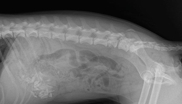
Figure 2: Lateral abdominal radiograph demonstrating a comminuted fracture affecting the cranial endplate of L6 with possible bone fragments within the vertebral canal and ventral to the remaining body of L6. On neurologic examination, the dog demonstrated non-ambulatory paraparesis. He had weak voluntary motor function in both pelvic limbs when supported. Myotatic reflexes in the pelvic limbs were intact. Tail movement and the perineal reflex were reduced. Further CT imaging was recommended.

Figure 3: Computed tomographic multiplanar reconstruction (MPR) sagittal (left) and transverse (right) plane images of the same dog in Figure 2 demonstrating foreshortening of the body of L6 due to a comminuted compression fracture of its cranial endplate. The cranial third of the vertebral body is completely and irregularly divided into several fragments of variable shape and size, many of which have displaced dorsally, invading the vertebral canal and causing marked compression of the spinal cord (up to 80 per cent of the diameter is compressed). A few large fragments are also seen ventral to the cranial aspect of the vertebra. Surgery was recommended, which included a mini-hemilaminectomy to decompress the spinal cord and stabilisation in the form of pins and bone cement.
Treatment
Treatment of vertebral fractures depends on neurologic status of the patient and the stability of the fracture. The goal of vertebral fracture management is to prevent further primary injury, realignment of the vertebral canal, stabilisation of the vertebral column and decompression of the spinal cord if indicated. Medial therapy is aimed at maintaining normal arterial oxygenation and systemic arterial blood pressure in an effort to minimise secondary spinal cord injury. The latter is achieved through use of intravenous fluid therapy and minimisation of anaesthesia-related hypotension as much as is feasible.
The use of corticosteroids as modulators of secondary spinal cord injury is controversial and there is currently no strong evidence to support their use. Furthermore, they are associated with complications such as gastrointestinal ulceration, melaena, diarrhoea, urinary tract infections, and in addition, their use precludes administration of non-steroidal anti-inflammatory drugs (NSAIDs). Surgery offers the best chance of realignment and stabilisation of the vertebral column and is indicated when there is paresis with intact nociception, instability, spinal cord compression, unmanageable pain, or worsening neurologic status.
Unstable vertebral fractures should be stabilised without delay. Indications for non-surgical management would include an ambulatory patient with minimal deficits and a stable fracture based on imaging. However, any patient that demonstrates neurologic deterioration or worsening of pain should be revaluated for possible surgical management. Non-surgical management consists of cage rest for at least four to six weeks, analgesia such as an NSAID, paracetamol +/- codeine, gabapentin, alone or in combination, and, in the case of cervical fractures, a possible neck brace.
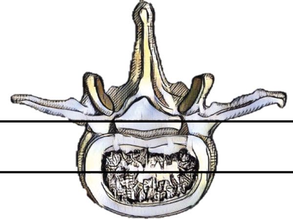
Figure 4: Three compartment model described by Denis in 1983. The dorsal compartment (above the upper black line) consists of the spinous process, vertebral laminae, articular processes, vertebral pedicles, and dorsal ligamentous complex consisting of the supraspinous ligament, interspinous ligament, joint capsule of the zygapophyseal (facet) joints, and ligamentum flavum. The middle compartment (within the upper and lower black lines) comprises the dorsal longitudinal ligament, dorsal annulus fibrosus, dorsal portion of the vertebral body. The ventral compartment (below the lower black line) consists of the remainder of the vertebral body, lateral and ventral portions of the annulus fibrosus, nucleus pulposus, and the ventral longitudinal ligament.
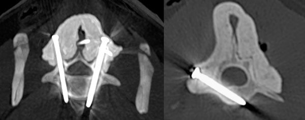
Figure 5: Immediate post-operative computed tomographic multiplanar reconstruction (MPR) transverse plane images of L7 (left) and L5 (right) of the same dog in Figures 2 and 3 demonstrating screw placement within the pedicles of L7 and one of two screws placed in the vertebral body of L5 (the other screw cannot be seen on this image slice).
Surgical stabilisation techniques
A variety of surgical stabilisation techniques have been described including use of plates and screws, external skeletal fixation (ESF), pins or screws and bone cement, tension band stabilisation, use of plastic spinous process (Lubra) plates, modified spinal segmental stabilisation, pedicle screw-rod fixation, and so forth. Several of these techniques such as tension band stabilisation, use of plastic Lubra plates, and modified spinal segmental stabilisation have become unpopular. These fixation methods can be broadly categorised into those that are inserted into the ventral half of the vertebral column such as pins or screws and bone cement, plates and screws, and ESF; and those that are applied at the level of the dorsal half of the vertebral column such as spinous process plating, tension band stabilisation and modified segmental spinal fixation.
Nowadays, the most common methods of vertebral stabilisation across all regions of the vertebral column include pins/screws in combination with bone cement (Figure 5), pedicle screw-rod fixation, and use of locking plates. The use of pins or screws in combination with bone cement or titanium rods offers a very versatile method of vertebral stabilisation in dogs. Instrumentation of the vertebral column in dogs is not without its challenges. A thorough knowledge of the anatomy of the relevant vertebrae is mandatory in order to ensure safe implant placement and avoid vertebral canal breach. In the thoracic spine, implants are typically placed into the pedicles which lie on either side of the vertebral foramen, whereas in the lumbar spine, implants are typically placed into the ventral-most portion of the pedicles and into the vertebral body. The use of pre-operative CT has really aided the surgeon in determining the optimal pilot hole entry point and trajectory.
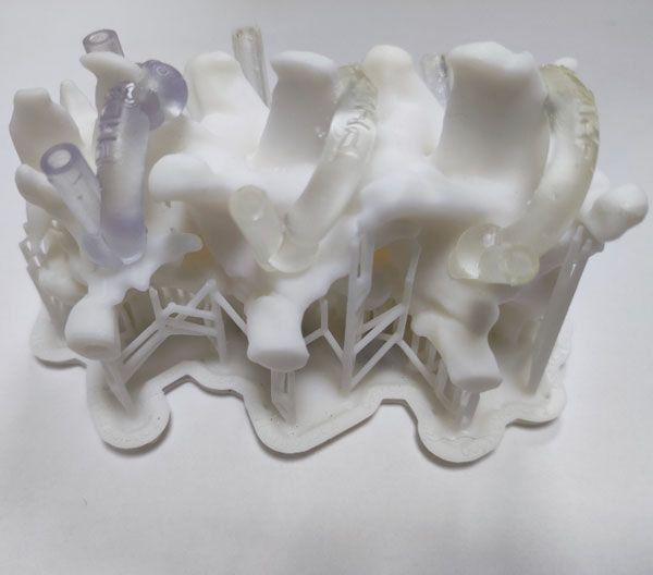
Figure 6: Three-dimensionally printed selected vertebrae and patient-specific drill guides.
Drill guides
A variety of techniques have been described to aid placement of implants into the canine vertebral column including the use of three-dimensionally printed patient-specific drill guides (Figure 6) based on pre-operative CT scans, the use of fluoroscopy, and a modification of the freehand insertion technique known as the pedicle-probing technique.
While 3D-printed drill guides are associated with very high accuracy and a low rate of vertical canal breach, they are expensive and require expertise to design and print. Furthermore, the hardware and software required to design and produce such guides are not universally available and external sourcing of guides can be associated with treatment delays. A good knowledge of the vertebral anatomy is still required in order to be able to place the guides correctly and to know when they are seated properly, with appropriate cleaning of any soft tissue interfering with the guide footprint. Recent literature has demonstrated the superior safety and accuracy of these guides over the traditional freehand technique.
A number of researchers have investigated the use of intraoperative fluoroscopy to guide placement of pins into the canine vertebral column. A recent publication by this author and collaborators from the United States and Europe demonstrated a high rate of accuracy associated with the pedicle probing technique. With this technique, the surgeon removes a small area of outer cortex (decortication) at the proposed site of implant insertion based on pre-operative CT scan, exposing the underlying cancellous bone. A blunt tipped probe or awl is inserted through this opening and advanced into the cancellous bone of the pedicle or vertebral body following the approximate trajectory based on pre-operative CT.
The principle of this technique is that the probe will follow the path of least resistance as it is advanced through the cancellous bone and establish the safe trajectory. The probe is then removed and the appropriate pilot hole drilled. The pedicle-probing technique also does not preclude the possibility of entering into the vertebral canal and good knowledge of the relevant anatomy is still required in order to be able to recognise anatomic landmarks and the appropriate implant entry (decortication) site.
One study led by this author involved comparing the safety and accuracy of pin placement in the thoracolumbar spine of canine cadavers using this technique versus 3D-printed drill guides. In that study, 112 positive profile pins were placed from T7 through L7, one left and one right. Post-operative CT was performed and pin tracts were graded by two blinded observers using a modified Zdichavsky classification. While both techniques had a high degree of accuracy, pins were placed faster using 3D-printed drill guides.
Further aspects of this research include investigating intraoperative techniques to detect vertebral canal breach prior to implant placement. One such initiative investigates the reliability of a dedicated probe instrument and image guided techniques to evaluate the integrity of pedicle drill tracts prior to implant placement. With non-image or 3D-printed guided techniques, the ability to determine a vertebral canal breach prior to implant placement would be particularly advantageous, especially in cases where pins or screws are combined with bone cement. If found to be reliable, these techniques could translate into decreased patient morbidity, reduced surgical interventions, decreased hospitalisation and reduced costs for clients.
Weh JM, Kraus KH. Vertebral fractures, luxations, and subluxations. In: S Johnston, K Tobias, eds. Veterinary Surgery: Small Animal. 2nd ed. Elsevier Saunders; 2018.
Bali MS, Lang J, Jaggy A, Spreng D, Doherr MG, Forterre F. Comparative study of vertebral fractures and luxations in dogs and cats. Vet Comp Orthop Traumatol. 2009;22(1):47-53.
Bruce CW, Brisson BA, Gyselinck K. Spinal fracture and luxation in dogs and cats: a retrospective evaluation of 95 cases. Vet Comp Orthop Traumatol. 2008;21(3):280-4.
Hettlich BF, Fosgate GT, Levine JM, Young BD, Kerwin SC, Walker M, Griffin J, Maierl J. Accuracy of conventional radiography and computed tomography in predicting implant position in relation to the vertebral canal in dogs. Vet Surg. 2010 Aug;39(6):680-7.
Denis F. The three column spine and its significance in the classification of acute thoracolumbar spinal injuries. Spine (Phila Pa 1976). 1983;8(8):817-31.
Mullins, RA, Espinel Ruperéz, J, Bleedorn, J, et al. Accuracy of pin placement in the canine thoracolumbar spine using a free-hand probing technique versus 3D-printed patient-specific drill guides: An ex-vivo study. Veterinary Surgery. 2023; 52(5): 648-660.


