UCD Research - April 2022
Does bone density in small animals decrease with age? A radiographic and CT study
Osteoporosis is a disorder of the skeleton characterised by bone loss and reduced bone density, which leads to an increased risk of fractures of the hips, vertebrae and wrists. It is a significant problem in older women, as the decreased levels of oestrogen which occur after menopause are linked to bone loss and increased porosity of bone. Considerable research has been conducted to determine if age-related bone loss occurs in domestic animals and whether animal models are suitable candidates for research on the development and progress of osteoporosis
In association with

H. Zhang BSc, School of Veterinary Medicine, UCD, Belfield, Dublin 4
N. Rosenberger MVB, School of Veterinary Medicine, UCD, Belfield, Dublin 4
A. Richardson BSc, BSc School of Veterinary Medicine, University of Liverpool, England.
D. Kilroy MVB, School of Veterinary Medicine, UCD, Belfield, Dublin 4
A. Kumar DVM, School of Veterinary Medicine, UCD, Belfield, Dublin 4
This research study was undertaken by a number of veterinary students who used radiographs and CT scans of dogs and cats to examine if skeletal changes occurring with age are indicators of osteoporosis in these species. As almost all female pets are sterilised at an early age, their low levels of oestrogen should influence bone density in later life.
Most of the research work was done online which meant that it was largely unaffected by the pandemic lockdowns.
Part one of study
The first part of the study examined digital radiographs of large breed (25-50kg) and giant breed (over 50kg) dogs to determine if anatomical variations in the sacrum and lumbar vertebrae contributed to lumbosacral pathologies. In addition, dissections of greyhound cadavers were conducted to examine in detail the anatomy of this region. In all cases (n=114), digital images were saved and analysed using ImageJ software which provided detailed data for study.
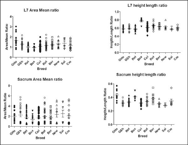
From this work, two biometric parameters were devised for both L7 and the Sacrum: Area-Mean Ratio (AMR) and Height-Length Ratio (HLR). The AMR is a measure of the relative bone density, while the HLR signifies the available surface area on the bone for a given unit length. Both parameters are inversely proportional to the characteristic that they represent – the lower the AMR, the higher the bone density, and the lower the HLR, the more rectangular the bone. Although the dogs in the study varied in terms of age, weight and sex, this was normalised during data analysis to make the parameters independent of any demographics. We concluded that significant differences in both anatomy and bone density occurred between the different groups of dogs (Cross breeds, large breeds, giant breeds1, Figure 1).

Figure 1. Range of measurements within selected breeds for Area-Mean Ratio (AMR) and Height-Length Ratio (HLR) of Lumbar vertebra 7 and the sacrum. Reproduced with permission from BEMS Reports1.
Part two of study
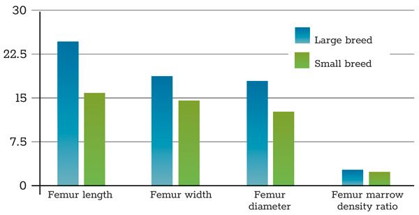
These findings stimulated a more specific study of CT images of the canine and feline skeleton to measure and analyse specific features (cross-sectional area of the femur; area of compact bone and extent of lumbar muscle mass). CT images of 14 dogs and six cats were obtained for examination. RadiAnt DICOM Viewer was used to read, measure and select suitable CT images and ImageJ software was used for further analysis. The width, height, area and pixel analysis (used to interpret bone density) were calculated for the fourth lumbar (L4) and fifth lumbar (L5) vertebrae, femoral heads, and pelvis in all subjects (Figure 2).
While there were no significant differences in the parameters measured, the stronger correlation between the density of L4 and L5 and the femoral head in cats compared to dogs indicates that cats may be more appropriate models for studying generalised osteoporosis, while dogs may be more suitable for localised disease.
Due to the small number of subjects examined, it was not possible to reach any definite conclusions on the extent of bone loss due to age. However, the work suggests that variables such as sex, breed type and gonadal hormone levels are unlikely to influence bone density2. Consequently, this work is continuing with a larger population of animals and it will include an examination of specific age-related changes in the skeleton.
Other factors need to be considered in deciding on suitable animal models for human conditions, specifically the very different reproductive cycles occurring in domestic animals. Dogs are classed as monoestrus and do not have a menstrual cycle; in addition, their levels of circulating oestrogens are significantly lower than in women. Furthermore, the fact that dogs and cats support their body weight on four limbs leads to different forces acting on the longitudinal vertebral column so biomechanical features of the quadruped and biped skeleton may play a part in the origin of this condition.
This research required few overheads as the images were provided from UCD veterinary hospital cases and free software (ImageJ and RadiAnt) were used for image analysis. The availability of enthusiastic students was the key driver of this work and their commitment and energy were greatly appreciated.
References
1. Rosenberger N, De Capitani E et al. Lumbosacral Biometrics and its Relevance to Patient-Specific Evaluation of Bone Mechanical Strength in Some Canine Breeds. Biology, Engineering, Medicine and Science Reports. 2019 Jul 1;5(1):05-9.
2. Zhang H, Kilroy D, Kumar AH. Do dogs get osteoporosis? A study of CT images of the canine pelvis. Irish Journal of Medical Science. 2021 Aug 1 (Vol. 190, No. SUPPL 4, pp. S131-S132).

Figure 2. Width, height, area and pixel analysis (used to interpret bone density) were calculated for the fourth lumbar (L4) and fifth lumbar (L5) vertebrae, femoral heads, and pelvis within selected breeds.
In association with

H. Zhang BSc, School of Veterinary Medicine, UCD, Belfield, Dublin 4
N. Rosenberger MVB, School of Veterinary Medicine, UCD, Belfield, Dublin 4
A. Richardson BSc, BSc School of Veterinary Medicine, University of Liverpool, England.
D. Kilroy MVB, School of Veterinary Medicine, UCD, Belfield, Dublin 4
A. Kumar DVM, School of Veterinary Medicine, UCD, Belfield, Dublin 4


