Coxofemoral Luxation in Cats: Aetiology, Diagnosis, and Management
Ronan A. Mullins, MVB DECVS PGDipUTL DVMS MRCVS, EBVS European specialist in small animal surgery, associate professor and discipline head of small animal surgery at University College Dublin, gives an overview of coxofemoral luxation in cats and its management
Coxofemoral luxation is commonly encountered in feline orthopaedic practice and is the most common joint luxation in this species. The condition involves the displacement of the femoral head from the acetabulum, leading to significant pain, lameness, and functional impairment. Understanding the aetiology, clinical presentation, diagnostic approaches, and treatment options for coxofemoral luxation is crucial for effective management and optimal outcomes. This article provides a comprehensive overview of coxofemoral luxation in cats, with an emphasis on surgical management and hip toggle stabilisation.
Anatomy of the coxofemoral joint
The coxofemoral joint is a simple, ball-and-socket, synovial joint, formed by the head of the femur articulating with the acetabulum. Primary stabilisers of the hip joint include the ligament of the head of the femur, the joint capsule, and the dorsal acetabular rim. No collateral ligament exits in the coxofemoral joint and the surrounding muscles allow a large range of motion. The ligament of the head of the femur originates from the acetabular fossa and inserts on the fovea capitis of the femoral head. Secondary stabilisers of the hip joint include the acetabular labrum, synovial fluid within the joint, periarticular muscles, and the transverse acetabular ligament.
Aetiology and Pathogenesis
Coxofemoral luxation in cats is primarily caused by trauma, with road traffic accidents being the most common cause. Other potential causes include kicks, fights, and falls from a height (Espinel Ruperez et al., 2021; LeFloch and Coronado, 2022). In a proportion of cases, the cause may not be known but trauma is suspected in the majority of these cases.
The pathogenesis of coxofemoral luxation involves disruption of the stabilising structures of the hip joint, including the joint capsule, ligament of the head of the femur, and surrounding musculature. Luxation occurs when at least two primary stabilisers are disrupted. The direction of the luxation, craniodorsal, caudoventral, or ventral depends on the nature and direction of the traumatic force (Cetinkaya et al., 2010; Ash et al., 2012; Espinel Ruperez et al., 2021; Pratesi et al., 2012; Knell et al., 2023). Craniodorsal luxation is the most common type, accounting for the vast majority of cases (Espinel Ruperez et al., 2021; Knell et al., 2023). With craniodorsal luxation, the femoral head is displaced cranially and dorsally relative to the acetabulum.
Clinical Presentation
Coxofemoral luxation is most commonly unilateral and predominantly affects young adult cats (Pratesi et al., 2012; Espinel Ruperez et al., 2021; Tamburo et al., 2022; Knell et al., 2023). Some studies demonstrate a preponderance of males (Pratesi et al., 2012; Tamburo et al., 2022), while in others, males and females are fairly equally represented (Espinel Ruperez et al., 2021; Knell et al., 2023). Domestic shorthairs are most commonly affected, likely because of their popularity in general (Pratesi et al., 2012; Espinel Ruperez et al., 2021; Knell et al., 2023). Although coxofemoral luxation has been reported in cats less than 10 months of age, trauma to the proximal femur in cats younger than this age is more likely to result in femoral capital physeal fracture.
Clinical signs of coxofemoral luxation in cats are often acute and include:
Lameness: Cats with acute coxofemoral luxation usually present with ambulatory non-weight bearing lameness on the affected pelvic limb if the luxation is unilateral and there are not concurrent orthopaedic/neurologic injuries (Knell et al., 2023). Bilateral non-weight bearing lameness (non-ambulatory) may be present if there is bilateral coxofemoral luxation and/or additional injuries in the contralateral limb.
Abnormal Limb Position: With craniodorsal luxation, the affected limb may appear shortened, externally rotated and adducted, whereas with caudoventral luxation, the limb may appear internally rotated and abducted. Additionally, an apparent slight increase in pelvic limb length may be identified when the limb is extended caudally. Conversely, a slight decrease in pelvic limb length may be identified when the limb is positioned cranially.
Diagnosis
Diagnosis of coxofemoral luxation involves a combination of orthopaedic examination findings and radiography.
Orthopaedic Examination
A thorough clinical examination is essential for diagnosis of coxofemoral luxation. In addition to lameness and abnormal limb position, orthopaedic examination findings associated with coxofemoral luxation can include:
Pain: The cat typically exhibits signs of pain when the hip is put through a range of motion.
Thumb displacement test: In a non-luxated hip joint, with placement of a thumb in the ischiatic notch between the greater trochanter and ischiatic tuberosity, external rotation of the femur will result in displacement of the thumb from the notch because the greater trochanter will occupy that space. In the case of a luxated hip, external rotation of the femur will not result in displacement of the thumb from the notch.
Loss of normal anatomic landmarks: The greater trochanter may be palpable in an abnormal position. With craniodorsal luxation, the greater trochanter will be palpable dorsal to an imaginary line between the craniodorsal ilium and the ischiatic tuberosity and equidistant between both. In a non-luxated joint, the greater trochanter is located ventral to this imaginary line and closer to the ischiatic tuberosity. With caudoventral luxation, there may be a decrease in the distance between the greater trochanter and the ischiatic tuberosity. Furthermore, the greater trochanter may be difficult to palpate.
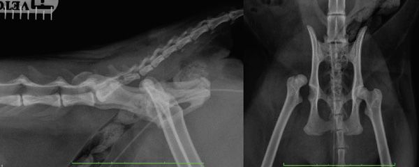
Figure 1: Preoperative lateral and ventrodorsal radiographs of a cat with right craniodorsal coxofemoral luxation.
Imaging
Radiography is the primary imaging modality used to confirm the diagnosis of coxofemoral luxation. Standard ventrodorsal and lateral views of the pelvis are obtained. Radiographic findings in coxofemoral luxation include:
Displacement of the Femoral Head: The femoral head is displaced from the acetabulum, either craniodorsally, caudoventrally, or in rare cases, ventrally (Figure 1).
Femoral Head and Acetabular Integrity: Assessment of the acetabulum and femoral head for fractures is critical as this will influence the treatment recommendation. Assessment for the presence of osteoarthritis should also be made.
Contralateral Hip Joint: Evaluation of the contralateral hip is important to rule out bilateral luxation or possibly any predisposing factors such as hip dysplasia.
In most cases, advanced imaging such as computed tomography is not necessary and should not replace the requirement for high quality, well-positioned radiographs.
Treatment
Treatment of coxofemoral luxation in cats can be broadly categorised into closed reduction, open reduction, and salvage procedures. The choice of treatment depends on factors such as the duration of the luxation, the presence of concurrent injuries such as fractures of the femoral head or acetabulum, the stability of the joint after closed reduction, cost, and the overall health and age of the cat.
Closed Reduction
Closed reduction is the least invasive option and involves manually manipulating the femoral head back into the acetabulum under general anaesthesia without surgical intervention. This can be attempted as the initial treatment for craniodorsal hip luxation but is associated with a high (~ 50%) re-luxation rate (LeFloch and Coronado, 2022). This high rate of failure may be related to the presence of remnants of the ligament of the head of the femur, joint capsule, and/or blood clots within the acetabulum preventing a stable reduction. Appropriate case selection is important and includes patients with craniodorsal luxation less than 5 days’ duration, without other orthopaedic injuries, and without concurrent fractures of the femoral head or acetabulum.
Procedure:
Goal: Bring the femoral head back into the acetabulum without iatrogenic cartilage injury.
General Anaesthesia: Closed reduction of coxofemoral luxation is performed under general anaesthesia to ensure muscle relaxation and pain control.
Positioning: Lateral recumbency with the affected limb uppermost.
Reduction Technique: With craniodorsal luxation, the femoral head is positioned on the dorsal aspect of the caudal ilium or dorsal to the acetabulum. The femoral head is disengaged from this abnormal position by grasping the hock and the stifle and externally rotating the pelvic limb. The limb is then pulled distally and caudally to align the femoral head over the acetabulum. Digital pressure applied to the greater trochanter during the procedure can be helpful in directing the femoral head into the acetabulum. With digital pressure applied on the greater trochanter, the femur is then internally rotated and abducted until the head snaps back into the acetabulum. A clunk should be palpated with successful reduction of the joint. In the case of caudoventral luxation, the femoral head is usually resting in the obturator foramen or cranial to it, hooked under the iliopectineal eminence. This type of luxation can be reduced directly into the acetabulum or can be converted to craniodorsal luxation and reduced as previously described. The femoral head is disengaged from the abnormal position by a gentle combination of traction and abduction of the limb. The femoral head is then elevated laterally and dorsally towards the acetabulum.
Post-Reduction Assessment: Successful reduction of the joint is confirmed with a lateral or oblique lateral (to separate left and right hemipelves) radiograph of the pelvis.
Post-Reduction Manipulation: Maintaining digital pressure on the greater trochanter and with internal rotation of the limb, the hip is put through a range of motion several times in an effort to displace any soft tissue within the acetabular fossa that may be preventing a tight and secure reduction. It is important that the hip is not externally rotated or abducted as this will most likely result in re-luxation.
Joint Immobilisation: Following successful reduction of craniodorsal luxation, the hip can be immobilised using a modified Ehmer sling to allow time for the joint capsule and supporting structures to heal. A hobbles can be used for caudoventral luxation to maintain the femoral head in the acetabulum. In one study, the median duration of bandage application was 12 days (LeFloch and Coronado, 2022). Application of an Ehmer sling does not guarantee that re-luxation will not occur. Such slings are often not well tolerated, they can be difficult to apply, can slip, and can cause skin wounds. Strict crate rest may therefore be more appropriate than relying on an Ehmer sling.
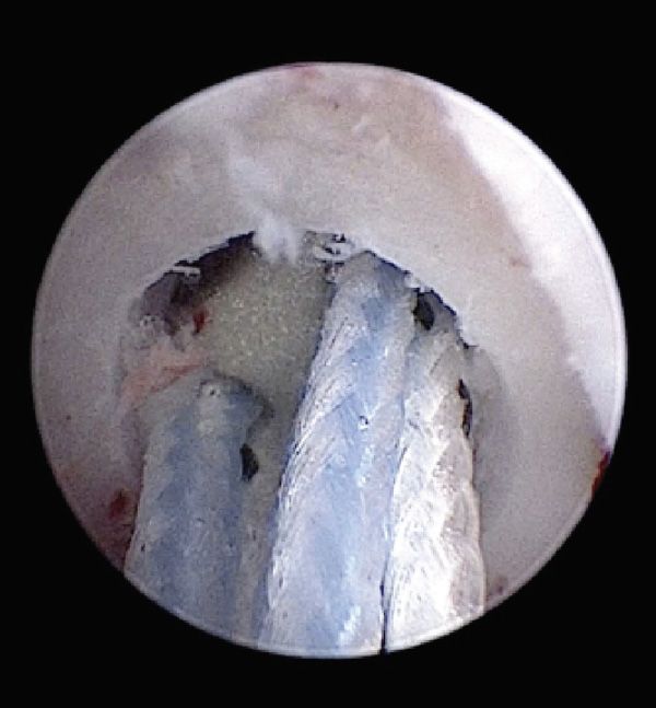
Figure 2: Arthroscopic image of the acetabular fossa in a cat demonstrating the metal toggle locked on the medial aspect of the acetabulum with attached strands of ultrahigh molecular weight polyethylene. Note the white articular cartilage at the top right of the image.
Open Reduction
Open reduction and internal fixation is indicated in cases where closed reduction is unsuccessful, the luxation is chronic, or there are concurrent fractures of the femoral head or acetabulum. Open reduction involves surgically exposing the hip joint and manually reducing the femoral head into the acetabulum, followed by stabilisation using various techniques. Several techniques have been described for the stabilisation of craniodorsal luxation in cats including but not limited to primary capsular repair, prosthetic capsule technique, ischioilial pinning, greater trochanter transposition, deep gluteal tenodesis, iliofemoral/rectus femoris suture, fascia lata loop stabilisation, transarticular stainless steel rope, transarticular pinning, and hip toggle stabilisation (HTS). Hip toggle stabilisation is currently one of the most commonly performed techniques to treat coxofemoral luxation in cats and will be focused on in this article. An advantage of this technique over some other techniques is that it permits early weight bearing on the affected limb, which may be particularly important in animals with contralateral limb injuries.
Hip Toggle Stabilisation (HTS):
Goal: The goal of HTS is to maintain coxofemoral reduction until joint capsular healing and periarticular fibrosis occur (Piermattei et al., 2016).
Surgical Approach: Both open and arthroscopic-assisted techniques have been described in cats. A craniolateral approach to the hip joint is the most commonly performed open approach. The approach to the hip joint can be made technically easier if the luxation can be reduced immediately preoperatively (Ash et al., 2012). The joint capsule is incised, and the femoral head is visualised. Small articular bone fragments from either the femoral head or dorsal acetabular rim may need to be removed (Pratesi et al., 2012). If such bone fragments were found to be very large, a salvage procedure may be required.
Surgical Procedure: The technique involves creation of two bone tunnels, one in the centre of the acetabular fossa and another in the femur extending from the fovea capitis to the level of the third trochanter. Remnants of the ligament of the head of the femur are first removed from the acetabulum. A metal toggle and attached strands of multifilament ultrahigh molecular weight polyethylene (eg, LigaFibaTM or TightRopeTM) or Nylon leader line is passed through the hole in the acetabular fossa and pulled up on sharply securing it on the medial aspect of the acetabulum (Figure 2) (Espinel Ruperez et al., 2021; Knell et al., 2023). Drilling of the femoral bone tunnel can be facilitated with use of a C-shaped aiming device. Without a guide, it is easier to externally rotate the femoral head and drill a bone tunnel from the fovea, through the femoral neck, exiting the lateral aspect of the femur distal to the greater trochanter. Free strands of polyethylene are then brought from proximal to distal through the femoral bone tunnel. The joint is reduced and traction applied on one of the strands to remove any slack. Strands are passed through a metal or plastic button and tied very tightly on the lateral aspect of the femur with the limb in a neutral position. An alternative to use of commercially available buttons involves creation of an additional bone tunnel, drilled from cranial to caudal through the lateral cortex of the proximal femur slightly proximal to the previously drilled femoral bone tunnel. One suture end is pulled through this additional drill hole and is tied to the other end of suture on the lateral side of the femur. Efforts should be made to close the joint capsule before routine closure as non-performance of capsulorrhaphy was associated with re-luxation in one study (Espinel Ruperez et al., 2021).
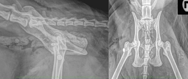
Figure 3: Postoperative lateral and ventrodorsal radiographs of a cat with right craniodorsal coxofemoral luxation following open reduction and hip toggle stabilisation. The metal toggle can be seen on the medial aspect of the acetabulum as well as the metal button on the lateral aspect of the femur. It is important that the femoral head is well reduced, without subluxation, that the femoral bone tunnel is contained within bone, and that the toggle and button are tight against bone.
Post-Reduction Assessment: Lateral and straight ventrodorsal radiographs of the pelvis with the pelvic limbs extended and parallel to one another should be obtained (Figure 3). The position of the metal toggle and the femoral drill tract should be assessed. A very mild degree of pelvic canal narrowing caused due to the presence of the toggle rod on the medial wall of the acetabulum may be present but is not a significant clinical problem in the author’s experience.
Possible postoperative complications: Reported postoperative complications in cats include re-luxation, implant-associated infection, development of osteoarthritis, an increase in the size of the femoral bone tunnel, wound-related complications (seroma, self-induced trauma to the surgical incision), lameness, transient sciatic neuropraxia (Knell et al., 2023; Tamburo et al., 2013; Pratesi et al., 2012). The overall re-luxation rate after HTS reported in the current literature in cats is 0-15.6% (Knell et al., 2023; Pratesi et al., 2012; Tamburro et al., 2022; Espinel Ruperez et al., 2021). Infection does not appear to be a common postoperative complication.
Postoperative Care: Typical postoperative care would include appropriate-sized crate rest for a period of 4-6 weeks (Espinel Ruperez et al., 2021). The author recommends ~ 4 weeks of crate rest followed by an additional 4 weeks of house rest. Appropriate perioperative analgesia (eg, NSAID) should also be provided as needed. Repeat orthopaedic assessment and radiographs of the pelvis should be performed at 6 weeks postoperatively. It has been reported that cats can have good clinical use of the limb despite the identification of re-luxation on follow-up radiographs (Pratesi et al., 2012).
Salvage Procedures
Salvage procedures are indicated for the treatment of recurrent femoral head luxation, concurrent severe fractures of the acetabulum or femoral head/neck, and possibly cases with significant hip osteoarthritis.
Femoral Head and Neck Excision (FHNE): FHNE, also known as femoral head ostectomy (FHO), involves the surgical removal of the femoral head and neck. This procedure eliminates bone-on-bone contact, reducing pain and allowing the formation of a pseudoarthrosis. Femoral head and neck excision should be considered if closed and open techniques are unsuccessful, if severe femoral head damage is present and total hip replacement is not an option, or if the owner is averse to the risk of re-luxation and additional surgery.
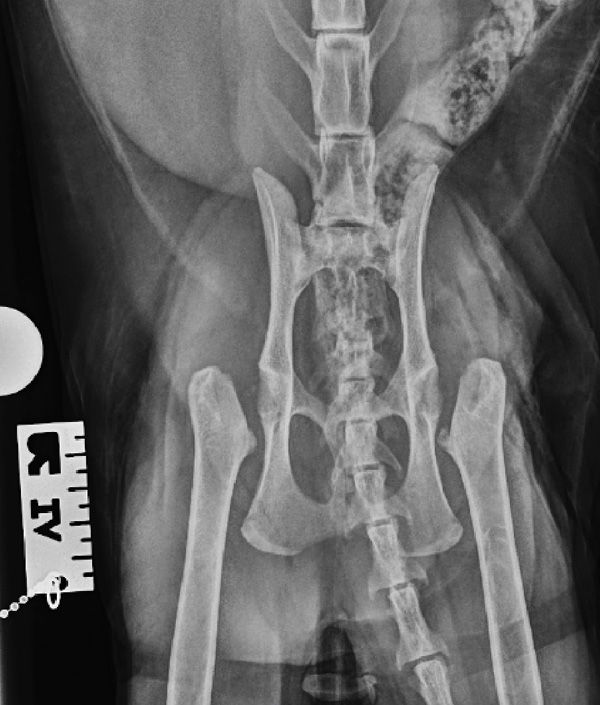
Figure 4: Staged bilateral femoral head and neck excision in a cat. Images courtesy of Dr Gareth Arthurs, Arthurs Veterinary Specialists, UK.
Procedure:
Surgical Approach: A craniolateral approach to the hip joint is used.
Surgical Procedure: Following incision in the joint capsule, the affected limb is externally rotated with the patella pointing toward the ceiling and maintained in that position during cutting of the bone. The osteotomy is performed with a sagittal saw from just medial to the greater trochanter to just proximal to the lesser trochanter (the site of attachment of the iliopsoas). Bone cut edges are wrasped and the site is lavaged. The joint capsule is closed, followed by routine closure of the remaining layers.
Postoperative Care: Postoperative care includes pain management and controlled physical therapy to encourage the formation of a functional pseudoarthrosis.
Total Hip Replacement (THR): Total hip replacement (Micro THR) may be used in cases of chronic luxation, re-luxation, severe osteoarthritis, or damage to the femoral head (Liska, 2010).
Procedure:
Preoperative Planning: Thorough preoperative planning using radiography are essential to appropriate implant sizing.
Surgical Approach: The hip joint is exposed through a craniolateral approach.
Surgical Procedure: The damaged femoral head is resected, and femoral and acetabular beds are prepared. Prosthetic implants are secured in place with bone cement.
Postoperative Care: Cats should be confined to a cage with avoidance of jumping. Appropriate perioperative analgesia (eg, NSAID) should also be provided as needed. Repeat orthopaedic examination and radiographs are obtained at ~ 6 weeks postoperatively, after which gradually increasing exercise activity is permitted, before returning to normal activity at ~ 12 weeks.
Prognosis
The prognosis for cats with coxofemoral luxation varies depending on the type and timing of treatment, the presence of concurrent injuries, and the overall health of the cat. Approximately 50% of cats that undergo closed reduction will experience re-luxation. Generally, cats treated promptly with open reduction without femoral head or acetabular fractures have a good prognosis, with many regaining normal or near-normal function. In one study in which long-term (>6 months) follow-up of 25 cats that underwent HTS was available, 92.0% demonstrated no lameness, while 8% had low to medium grade lameness (Espinel Ruperez et al., 2021). Similar findings were reported in another study in which all cats showed either minimal or no lameness at a mean of 13 months postoperatively (Knell et al., 2023). Finally, in a study by Tamburo and others (2022), final outcome at 12 months postoperatively was considered excellent in 76% cats and good in the remaining 24%.
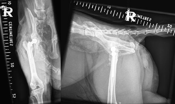
Figure 5: Right-sided cemented total hip replacement in a cat. Images courtesy of Dr Gareth Arthurs, Arthurs Veterinary Specialists, UK.
Ash K, Rosselli D, Danielski A, Farrell M, Hamilton M, Fitzpatrick N. Correction of craniodorsal coxofemoral luxation in cats and small breed dogs using a modified Knowles technique with the braided polyblend TightRope systems. Vet Comp Orthop Traumatol. 2012;25(1):54-60.
Cetinkaya MA, Olcay B. Modified Knowles toggle pin technique with nylon monofilament suture material for treatment of two caudoventral hip luxation cases. Vet Comp Orthop Traumatol. 2010;23(2):114-118.
Espinel Rupérez J, Arthurs GI, Hewit A, et al. Complications and outcomes of cats with coxofemoral luxation treated with hip toggle stabilization using ultrahigh-molecular-weight-polyethylene or nylon (2009-2018): 48 cats. Vet Surg. 2021;50(5):1042-1053.
Knell SC, Longo F, Wolfer N, Schmierer PA, Hermann A, Pozzi A. Outcome and Complications following Stabilization of Coxofemoral Luxations in Cats Using a Modified Hip Toggle Stabilization-A Retrospective Multicentre Study. Vet Comp Orthop Traumatol. 2023;36(4):218-224.
LeFloch MD, Coronado GS. Outcome of coxofemoral luxation treated with closed reduction in 51 cats. J Feline Med Surg. 2022;24(8):709-714.
Liska WD. Micro total hip replacement for dogs and cats: surgical technique and outcomes. Vet Surg. 2010;39:797-810.
Piermattei D, Flo G, DeCamp C. The hip joint. Brinker, Piermattei, and Flo’s Handbook of Small Animal Orthopedics and Fracture Repair. 5th ed. St. Louis, MO: Elsevier; 2016: 468-480.
Pratesi A, Grierson J, Moores AP. Toggle rod stabilisation of coxofemoral luxation in 14 cats. J Small Anim Pract. 2012;53(5):260-266.
Tamburo R, Pratesi A, Carli F, Collivignarelli F, Bianchi A, Paolini A, Ilaria F, De Bonis A, Vignoli M. Traumatic Coxofemoral Luxation in Cats Treated with Hip-Toggle Stabilization Using the Mini Tightrope® Fixation System. Acta Veterinaria-Beograd. 2022;72(3),300-308.
1. What is the most common direction of coxofemoral luxation in cats?
A. Caudoventral
B. Ventral
C. Craniodorsal
D. Cranioventral
2. Primary stabilisers of the hip joint in cats include which of the following?
A. Ligament of the head of the femur, joint capsule, dorsal acetabular rim
B. Medial and lateral coxofemoral collateral ligaments
C. Acetabular labrum, synovial fluid, and periarticular muscles
D. Annular and transverse acetabular ligaments
3. Cats with craniodorsal hip luxation may carry the affected limb in which of the following positions?
A. Externally rotated and adducted
B. Externally rotated and abducted
C. Internally rotated and adducted
D. Internally rotated and abducted
4. According to a recent publication, what is the reported rate of re-luxation after closed reduction of coxofemoral luxation in cats?
A. 30%
B. 40%
C. 50%
D. 60%
5. With craniodorsal luxation, which of the following orthopaedic examination findings would be most likely?
A. The greater trochanter will be palpable ventral to an imaginary line between the craniodorsal ilium and the ischiatic tuberosity and closer to the craniodorsal ilium
B. The greater trochanter will be palpable ventral to an imaginary line between the craniodorsal ilium and the ischiatic tuberosity and closer to the ischiatic tuberosity
C. The greater trochanter will be palpable dorsal to an imaginary line between the craniodorsal ilium and the ischiatic tuberosity and closer to the craniodorsal ilium
D. The greater trochanter will be palpable dorsal to an imaginary line between the craniodorsal ilium and the ischiatic tuberosity and equidistant between both
ANSWERS: 1C; 2A; 3A; 4C; 5D


