


Dermatology treatment for cats and dogs: multi-modal therapy care
Feline and canine dermatology is an excellent example of where practical and affordable non-pharmacy approaches can be a determinant of treatment success. Dr Aine Seavers MVB MRCVS, outlines some non-pharmacy treatment strategies for a range of dermatological conditions
As the global veterinary profession moves back to a more nuanced individualised treatment approach, it is timely to consider how successful treatment outcomes can be achieved without reaching for drugs as much or, perhaps, at all.
There are a number of things that can assist healing without polypharmacy (the use of multiple prescription drugs), such as: mechanical decontamination and debridement; protective and healing clothing, such as sun-vests and elbow shields; hydro-jet therapy; laser/photobiomodulation therapy; and topical, rather than oral, chemotherapy options.
Taking the clinical history of the patient is the most important step of all in detecting and removing the primary culprit before any veterinary treatment commences. A good clinical history can avoid the wasteful use of a drug, save the client from unnecessary cost, and save the pet from a possible adverse drug response and a lost opportunity to have their skin condition cured rather than controlled. In this article, I present six examples of treatments for dermatological conditions including some examples of non-pharmacy solutions.
1. Applying Imiquimod topically for SCC or MSCCIS in cats
Imiquimod 5% cream (Aldara) is a novel immune response modifier (IRM) shown to have indirect antiviral and antitumour effects. Imiquimod enhances both innate and cell-mediated immunity. Aldara 5% is labelled for use in actinic keratosis, superficial basal cell carcinoma, and external genital warts in humans.
Multicentric squamous cell carcinoma in situ (MSCCIS) is a recently described variant of squamous cell carcinoma (SCC) in cats. MSCCIS is also referred to as a ‘Bowen-like’ disease because of its histologic resemblance to Bowen’s disease in humans. Aldara has been reported as a successful treatment for Bowen’s disease in humans.
How to use Aldara/Imiquimod in early SCC presentations
If the lesions are overly crusty, first clean up the sites. This can be done during the consult, if the cat tolerates the cleaning and if the lesions are not overly friable. Otherwise, admit to lightly sedate, then debride.
Once clean, ‘paint’ the Aldara onto the lesions. I requested that the owner in this case use tiny cosmetic cotton make-up buds to paint the cream onto the lesion as this seemed to stop the owner from being heavy-handed in application of the cream. However, one can just rub it straight onto the lesion. Gloves must always be worn by the applier.
Application frequency for cats
Apply once daily, three days on and four days off. Expect to see inflammation in the treatment area as this is an indicator that Aldara is working. However, in the occasional case where the inflammation is extreme, have the owner demonstrate how they apply the cream.
If the owner’s manner of application is appropriate, stop treatment for 48 hours, then resume every second day. If the ensuing degree of inflammation is still not tolerated by the cat, then move to a new treatment option. If the drug is tolerated, then the treatment continues for six to 16 weeks weeks until the lesions resolve and dry up.
Aldara is available as a box of either six or 12 sachets. Depending on how large the target area is, it is possible to get one to two weeks out of each sachet if the owner seals the packet back up each time and keeps the product airtight.
Do your own research as to purchaser options before prescribing Aldara. Prices to clients vary hugely depending on the human pharmacy where the product is bought, so make sure to investigate the purchase cost well in advance.
In addition, owners should be advised on how to stop their cats from sunbathing indoors and outdoors. In Australia, we see an increase in the number and severity of SCC cases at the end of the winter as the colder weather means cats can/will remain longer in the available winter sunlight, thus increasing the actinic damage risk. For further tips to help cats reduce their time sunbathing, see article, “The heat is on: the feline TNZ” in Veterinary Ireland Journal, Vol 13, No. 4, pages 200-201.
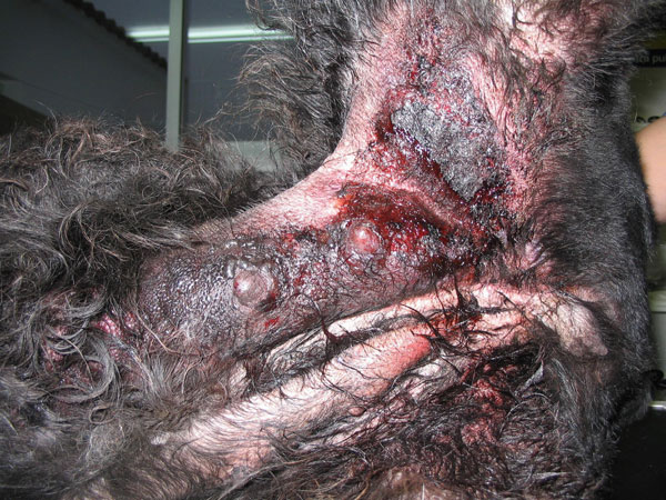
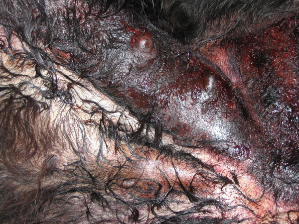
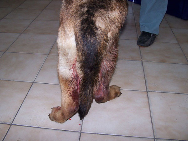
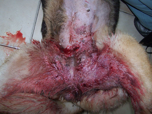
Figures 1-4: Examples of inflammatory malignant metastatic mammary adenocarcinoma (IMMMAC). Photos 1 and 2, courtesy of Dr Pedro Ginel. Photos 3 and 4, courtesy of Dr Yovko Haralanov.
2. IMMMAC in dogs
When it comes to sheer pain and agony, very few conditions come close to inflammatory malignant metastatic mammary adenocarcinoma (IMMMAC) (see Figures 1 to 4 on facing page). The sooner these patients are given the last gift an owner can offer – a gentle death via euthanasia – the better for these poor dogs.
IMMMAC is an aggressive form of mammary tumour seen in dogs and humans. In dogs, the condition often causes mammary glands to become swollen and painful.
The swelling can be focal or diffuse and involve either a single mammary chain or all mammary glands.
Harvesting a biopsy early on in the presentation is the preferred option to ensure you and the client make a fully informed decision from day one. However, where such a gold-standard option is not feasible, a presumptive guarded diagnosis based primarily on pattern recognition is also contextually valid. It can be difficult to get a differential diagnosis list going in one’s mind; however, once you have seen your first IMMMAC case, you will recognise it the next time.
The heat and erythema are 10/10. These cases are the stuff of nightmares; the experience is slow to leave your mind, including the vocalisation and the shivering as the animals writhe on the consult table. This is no time to be heroic but it is time to stop the suffering. Poor appetite, loss of condition, generalised weakness, and oedema of one or both hind legs may also be present.
Treatment
Despite one paper showing the life-extending effectiveness of piroxicam in a group of these patients, my experience, and that of over 1,500 vets I canvassed on this condition, is that by the time they are presented to you, it is too late.
Occasionally, the patient will get a short reprieve of one to two weeks on antibiotics and non-steroidal anti-inflammatory drugs (NSAIDs), but the condition roars back with a vengeance meaning all you have done is set the patient up for even more agony. This presentation is also one of these occasions where there is minimal cross-over efficacy of meloxicam to piroxicam in suppressing the neoplastic disease.
Do not attempt to surgically mammary-strip these patients. This will only put the animal through the stress of the anaesthetic and the surgical pain with absolutely no effect on the progress of the IMMMAC. Radiation therapy and targeted monoclonal antibody (MAB) therapy in humans are part of an extensive palliative multimodal therapy regime for this condition.
The next time you are presented with a female dog displaying massive erythema in the mammary area, with 10/10 heat, and, in most cases, hindlimb oedema (often bilateral), you must rule out IMMMAC before commencing treatment or surgery.

Figure 5: Acute moist dermatitis/hot spot/pyotraumatic dermatitis. Photo courtesy of Dr Candace Sousa.
3. Shining lasers on ‘hot spot’/pyotraumatic dermatitis/acute moist dermatitis
There is an ever-increasing emphasis on the need to minimise antibiotics use in order to avoid disturbing the balance of the skin microbiome. The more presentations that are highlighted as valid for non-drug treatment options, the more we can move away from blanket destruction of the microbiome by polypharmacy and instead support the body to recalibrate the particular condition back to a healthier balance.
Pyotraumatic dermatitis is one such presentation. With this condition, I am a firm believer in shaving, debriding, and cleaning up these hot-spot areas before starting any pharmacy. Too many dogs present as repeat sufferers without having had proper prevention or decontamination measures carried out previously. This leaves owners expecting their pets to undergo heavy-duty medication for a protracted period.
In addition to gentle debridement, consider adding laser/photobiomodulation (PBM) therapy to these skin presentations. Don’t just treat the affected area with the laser; treat the peripheral tissue also to assist in the resolution of inflammation. Treating beyond the lesion boundaries decreases the levels of inflammatory tissue markers swamping the lesion and helps prevent the local nociceptors from firing furiously and worsening the self-inflected trauma and overall visual presentation.
In applying laser/PBM therapy to these presentations, reliance on the client’s home compliance efforts is reduced. The PBM therapy will assist with a reduction in pain and self-trauma and hence facilitate a speedier recovery for the patient.
Using PBM therapy could also reduce the number of pyotraumatic dermatitis cases needing antibiotic therapy as part of their hot-spot treatment. I discussed this topic of hotspot and PBM with a group of dermatology vets. In the course of the discussion, a valid point was made in relation to the Golden Retriever breed. One vet pointed out that the AMD in this breed - particularly on the face/neck/head - may not be pyotraumatic, but rather a manifestation of superficial bacterial pyodema.
This is an important point about specific breeds and the bacterial aspect of the condition as some presentations will need antibiosis.
When using photonic therapy, it’s important to remember that different PBM therapy machines use different wavelengths and that, in turn, those wavelengths each have different effects on bacteria, viruses, etc. Some wavelengths such as blue light, could indiscriminately sterilise the entire treatment area – a result that one rarely desires.
The most commonly used veterinary NIR Class 4 (810-980nm) machines don’t directly kill bacteria. The main target of the NIR light is mitochondria, which bacteria don’t contain. Therefore, one is less concerned about wiping out the microbiome with 800-910 wavelengths, while still getting an antibacterial effect due to the body’s improved internal intrinsic signalling.
PBM therapy won’t replace antibiotic therapy where it is needed, but it will eliminate a host of cases where antibiotics might have been unnecessarily dispensed.
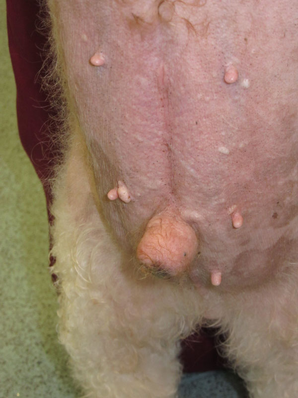
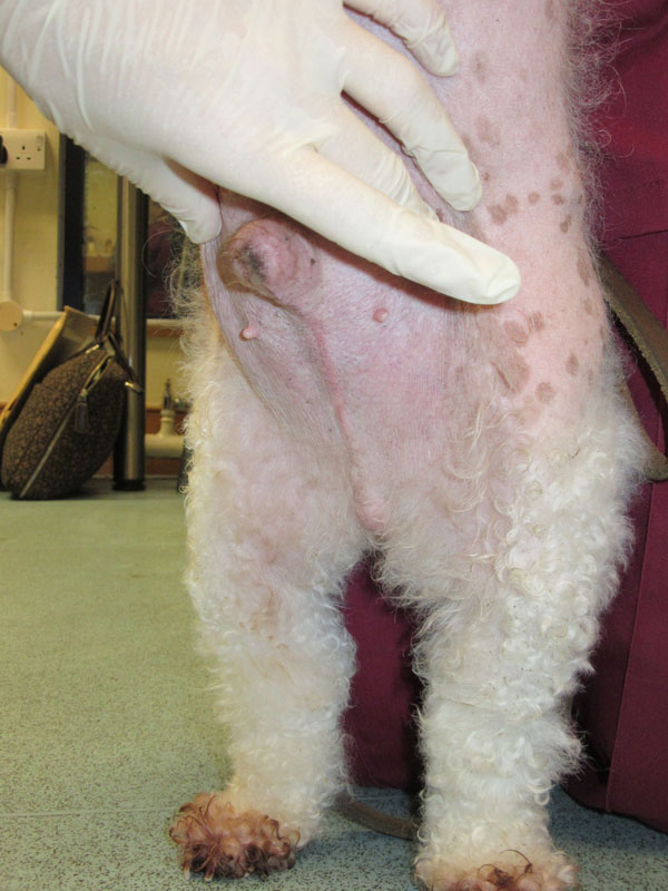
Figures 6 and 7: Examples of hormone replacement cream-induced hypertrophy. Photos courtesy of Dr Victoria Robinson.
4. Hormone replacement creams – cover up!
Vets must be alert to the unintended side effects of human hormone replacement therapy on our veterinary patients. The increased prescribing of topical hormone treatments for a wide range of people of varying ages – not exclusively menopausal women – is having unintended wider medical consequences for pets living with owners receiving this topical hormone therapy.
The condition was first reported back in 2009, yet I find it continues to be initially misdiagnosed. In 2009, the marketer of the spray-on female topical oestradiol product (Evamist) notified the US Food and Drug Administration (FDA) of eight reports of children, and several of animals, being inadvertently exposed to the topical hormone treatment. In humans, mammary development occurred in young males and females several weeks or months after older women in the family began topical hormone replacement therapy. Symptoms abated after the women either ceased using the product or took steps to avoid exposing others.
Hormone replacement treatments, such as lotions, gels, or sprays, were normally applied to the arms – especially inner elbows and wrists – or legs. Unfortunately, that meant when the humans held, petted, or snuggled up with their dogs, unforeseen amounts of the drug were then absorbed by the pets. Additional exposure occurred when pets licked their owner’s medicated skin or accessed and ate the discarded transdermal patch or tubes.
When vets were first presented with these cases, they were perplexed, and either diagnosed ovarian remnant syndrome post desexing, or other endogenous hormone aberrations, or adrenal disease. The dogs would often yelp as if in pain, suggesting internal disease concerns. Desexed male dogs presented with enlarged mammary glands and abnormally small penises. The ventrum skin often had the texture of plastic. Alopecia was also often present. Desexed young female puppies presented to vets with markedly swollen vulvas as if in oestrus.
In cats, the signs resemble classic ‘in heat’ behaviour: calling, rolling, etc.
Some young animals, who were accidentally exposed to their owners’ hormone replacement therapy for over 12 months, then took another year to recover even after all exposure had ceased.
Young children in the same family may also be contaminated by the hormone creams and have behavioural issues, so it is worthwhile being alert to any comments clients make about concerns with other family members.
Today, 15 years after the first reported incidents, we continue as frontline vets to see these cases, often at second-opinion stage. In many of these instances, the client can become angry, thinking that the first vet did not neuter or spay their pet correctly. Sometimes a client reveals that they had not told the first vet that they, the owner, were using hormone cream. It is so important to communicate to the client the need for them to fully inform you of any exposure to hormone creams, in order to prevent the pet from undergoing invasive and expensive investigations.
Prevention is simple
Users of topical hormone products simply need to wear gloves to apply the product to areas that can be then covered with clothing. Vets must ask the client about anyone in the in-contact family who may be using hormone replacement therapy. A detailed history-taking of any unusual presentation is usually the key to finding the correct trigger agent.

Figure 8: Hygroma harness. Photo courtesy of Kelly Vella.
5. Curing elbow hygromas with cloth and/or light
I have found elbow hygromas in dogs one of the hardest conditions to heal, and the prevention of subsequent relapse was equally difficult. These challenges were particularly evident with elderly dogs that spend a lot of their day sleeping, recumbent on those worsening pressure sores. However, the discovery of an elbow harness (from DogLeggs) to treat and heal hygromas has been a game changer. Using just the harness alone, even the worst lesions heal rapidly. Once healed, the harness prevents new pressure sores from forming.
Nowadays, given access to a Class 4 laser/PBM machine, I would use that as well to hasten the speed of healing of the ulcerated areas in cases where the owner is awaiting the delivery of the harness.
The dog in Figure 8 was a 14-year-old Staffordshire Bull Terrier with weeping pressure sores and trophic ulceration on her forelimbs. Without any parenteral medication, her elbows began to heal within days of the dog wearing the hygroma harness.
The harness also appeared to give support and security to this old dog; she appeared to feel safer with the harness as it also supported her and prevented her front legs from slipping out sideways as she stood up. I have since used the harness in other geriatric dogs, especially giant breed dogs, to give them stability as they rise.

6. Fournier’s gangrene
The cat in Figure 10 presented to a colleague some years ago with severe diarrhoea and prolapse of a small amount of rectal mucosa. She initially responded well to treatment but developed extensive necrotising dermatitis, assumed to be secondary to gastrointestinal flora invasion across the damaged rectal mucous membranes and into the perineal tissues. In a non-specific history case, fever plus pain disproportionate to the appearance of the affected area should alert vets to the possibility that this is not your average cat abscess in the perianal, perineal and scrotal area.
Necrotising fasciitis is a fulminant, rapidly progressive, potentially life-threatening, bacterial infection of the fascial and subcutaneous tissue. In human medicine, there is a variant of necrotising fasciitis called Fournier’s gangrene.
It is defined as a necrotising soft tissue infection of the genital and anorectal regions characterised by tissue necrosis (cellulitis, fasciitis, and myositis), rapid progression, lack of frank suppuration, and severe systemic toxicity.
Fournier’s gangrene has been reported in cats and dogs. Biopsy for histopathology and culture is recommended. If the histopathology is consistent with necrotising fasciitis and the tissue samples are positive on culture, treatment involves supportive care (wound care, management of the systemic inflammatory response, analgesia) and antimicrobial therapy selected via culture and sensitivity.
Treatment and prognosis
Use a local anaesthetic and small scalpel-blade incision. A positive ‘finger test’ (i.e., easy-to-dissect subcutaneous tissue from the deep fascia tissues by blunt dissection using surgeon’s index finger) is a key marker in humans. A positive finger test means immediate and complete surgical intervention and debridement of the necrotic tissue, often requiring multiple surgical procedures. Surgical exploration is often performed every 24 to 48 hours on human cases and debridement is continued until the infection is completely cleared.
Another frequent surgical finding is the presence of copious, exudative, thin, malodorous fluid resembling ‘murky dishwater’. The fluid contains bacteria and leukocytes. Poor antibiotic delivery to the affected area and the continued production of bacterial toxins makes medical therapy in the absence of surgical debridement useless. Amputation of a limb is sometimes required in order to achieve complete excision of affected tissue.
Initial broad-spectrum antibiotic therapy is recommended while awaiting culture results. The triad of penicillin, an aminoglycoside, and metronidazole or clindamycin is the most frequently advocated antibiotic regime in humans. Inclusion of a clindamycin showed a trend toward better survival. Fluroquinolones need to be given at maximal concentration, a once-daily full dose is preferable to a split daily dose. Haemodynamic support, nutritional support, and analgesia are also essential.
Hyperbaric oxygen studies on this condition showed mixed results from this therapy.
Newer modalities of ulcer/wound management include: laser/PBM therapy, hydro jet debridement, and platelet rich plasma. These modalities can be used to augment the existing principles of management and to reduce the morbidity and mortality associated with the condition.

Figure 10: Suspected Fournier’s gangrene. The cat in the above image made a full recovery over a four-week period. Photo courtesy of Dr S. McMillan.
Suggested reading
- Inflammatory mammary carcinoma in 12 dogs: Clinical features, cyclooxygenase-2 expression, and response to piroxicam treatment. Can Vet J. 2009 May; 50(5): 506-510.
- Histological, immunohistological, and ultrastructural description of vasculogenic mimicry in canine mammary cancer. Vet Pathol. 2010 Mar;47(2):265-74. Epub 2009 Dec 31.
- Diagnostic Reasoning Strategies and Diagnostic Success. Med Educ 2003;37: 695-703.
- Multicentric squamous cell carcinoma in situ resembling Bowen’s disease in cats. Veterinary Pathology 1993; 30: 535-543.
- Tumours of the skin and subcutaneous tissues. In: Withrow & MacEwen’s Small Animal Clinical Oncology, 4th ed., SJ Withrow and DM Vail, eds., New York, Saunders Elsevier, 2007: 375-40.
- Hyperandrogenism after transfer of topical gel: case report and review of published and unpublished studies. Human Reproduction 200924:425-428
- Sexual precocity in a 4-year-old boy. BMJ 2010;340:c2319
- Role of newer technologies in wound bed preparation in Fournier’s gangrene . Plastics and Aesthetic Research 2018. June 2018; 5(6):20.
- Necrotizing Fasciitis: A Review. JAAHA 2005; 41: 104-109.
- Fitzpatrick’s Dermatology in General Medicine. 6th ed. New York: McGraw Hill Medical Publishing Division; 2003, p1102.
- Fournier’s gangrene in a cat. Journal of Veterinary Emergency and Critical Care. 08 February 2010. https://doi.org/10.1111/j.1476-4431.2009.00453.x.
- A case of necrotizing fasciitis with septic shock in a cat caused by Acinetobacter baumannii. Vet Dermatol 2007; 18: 432–438.
- Necrotizing fasciitis in a dog. J Vet Emerg Crit Care 2001; 11: 299–305.
1. Which of the following statements is not correct?
A. IMMMAC is an aggressive form of mammary tumour seen in dogs and humans
B. Dogs with inflammatory mammary carcinoma are often painful with swollen mammary glands
C. The swelling can be focal or diffuse and involve either a single mammary chain or all mammary glands
D. NSAIDs, antibiosis and radiation therapy are curative
2. Which of the following statements are correct?
A. Imiquimod 5% cream (Aldara) is a novel immune response modifier with indirect antiviral and antitumour effects
B. Aldara 5% is labelled for use in actinic keratosis, superficial basal cell carcinoma, and external genital warts in humans
C. MSCCIS is also referred to as a ‘Bowen-like’ disease in cats because of its histologic resemblance to Bowen’s disease in humans
D. One should not expect to see any inflammation in the treatment area after Aldara application
3. In pyotraumatic dermatitis, which treatment options can one consider?
A. Gentle debridement
B. Topical and oral anti-inflammatory and antibiotic therapy
C. Adding Laser/PBM therapy to the affected area and the peripheral tissue
D. All of the above
4. In relation to human hormone replacement creams, which of the following statements is incorrect?
A. Topical hormone supplement is available for a wide range of people of varying ages, not exclusively menopausal women
B. Animals are exposed to the hormone indirectly via skin contact with the human skin, by licking, and by actual ingestion of discarded transdermal tubes or patches
C. Young animals fully recover in a matter of days once all exposure has ceased
D. Desexed male dogs present with enlarged mammary glands, abnormally small genitalia, ventrum skin often has the texture of plastic and alopecia is often present
E. Users of topical hormone products need to wear gloves to apply the product to areas that can then be covered with clothing
5. In relation to necrotising fasciitis, which of the following statements is correct?
A. Fournier’s gangrene is defined as a necrotising soft tissue infection of the genital and anorectal regions characterised by tissue necrosis (cellulitis, fasciitis, and myositis), rapid progression, lack of frank suppuration and severe systemic toxicity
B. Fournier’s gangrene has been reported in cats and dogs
C. Biopsy for histopathology and culture is recommended
D. If the histopathology is consistent with necrotising fasciitis and the tissue samples are positive on culture, treatment involves supportive care, i.e., advanced multi-modal wound care therapies, management of the systemic inflammatory response, analgesia and antimicrobial therapy selected via culture and susceptibility
E. All of the above
ANSWERS: 1D; 2A, B, and C; 3D; 4C; 5E