Feline chronic gingivostomatitis and caudal feline malocclusion: treating each in first opinion practice
Nora Schwitzer DiplTier PhD GPCert(SADen&OS) MRCVS, MyVet Lucan, Co Dublin explores how to distinguish between feline chronic gingivostomatitis (FCGS) and caudal feline malocclusion and how to treat each of the two diseases in first opinion practice
FCGS and caudal malocclusion in cats are two very different diseases which both present with inflammatory reactions in the mouth. Both diseases are painful and both diseases can look similar at their first presentation. The prognosis of FCGS and caudal malocclusion however is very different. FCGS is a debilitating and frustrating disease which is difficult to treat and has a high proportion of refractory cases. Caudal malocclusion will resolve after the traumatic occlusion is corrected and only ‘re-appears’ with other concurrent dental disease later in life. In the article below I will describe each disease individually and give an update on the current treatment options available.
FCGS
FCGS is a painful, chronic debilitating disease in cats that lasts months to years. The cause of FCGS remains unknown but seems to be the manifestation of an inappropriate immune response to antigenic stimulation. It is therefore classified as an immune mediated disease. A recent study from Fried et al (2021) shows a strong association between FCV and FCGS with 21 out of 23 cats being affected but none of the control group cats. Additionally puma feline foamy virus was detected in cats that were refractory to treatment. Peralta et al (2019) could furthermore highlight that multi-cat environments seem to be an important contributing factor. Cats with FCGS were significantly more likely to come from shared households, and cats in shared households had significantly increased odds of FCGS compared with those from single-cat households.
Patients that present with FCGS usually suffer from weight loss, severe halitosis, are often depressed, have matted fur and are usually head shy.
Given the chronic pain FCGS patients are suffering from, a conscious dental exam can be challenging.
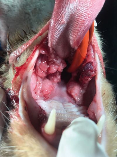
If it is possible to assess the cat’s mouth, the examiner can see erosive and/or proliferative inflammatory mucosal lesions lateral to the palatoglossal folds in the back of the mouth (Lommer 2013, see Figure 1). These inflammatory mucosal lesions lateral of the palatoglossal folds are the main characteristic or cardinal diagnostic features of FCGS. They can be ulcerative or proliferative and are always of bright red to dark red colour with a distinct smell. They 'look and smell sore’. Recent research has shown that FCGS is also always accompanied by periodontitis (Lee et al, 2020). Additionally, external inflammatory root resorption is common and makes treatment more challenging.
A diagnosis is supported by the attachment loss visible on radiographs, potential inflammatory root resorption and histopathology findings – lesions are infiltrated by lymphocytes and plasma cells, with fewer neutrophils, macrophage-like cells, and mast cells. For more information see Rolim et al (2017).
Patients with FCGS also seem to suffer from concurrent esophagitis. In the day-to-day treatment in clinical practice, this part of the disease is probably least well-managed and may aggravate the existing condition and contribute to inappetence, Kouki et al (2017).
FCGS is extremely painful and all patients should be offered immediate pain relief (depending on the patient and their comorbidities, NSAIDs, Gabapentin and opioids are options).
The current standard of care involves dental extractions of at least all premolar and molar teeth, with or without medical management, rather then medical therapy alone, Lee et al (2020). Premolar and molar extractions in these patients can be challenging due to the inflamed and frail gingiva and the nature of brittle, long, delicate and, in places, ankylosed cat tooth roots. However decapitations, tooth root remnants and extractions of only ‘individual particularly bad’ premolars or molars should be avoided, since these operations make future extractions (in a mouth that now contains inflamed tissue and scar tissue) even more challenging.
Additionally, owners need to be aware that only about one-third of all FCGS cases are much improved after the extraction of premolar and molar teeth or full mouth extractions. Many patients will depend on life-long medication afterwards.
Cyclosporin had the most promising results with 77 per cent of all cats treated improving (Lommer 2013) whereas corticosteroids at 1mg/kg only led to an improvement of one-third of the patients in the study, Hennet et al (2011). For “An overview regarding therapeutic management of FCGS: A systematic review of the literature”; please see Winer et al (2016).
Future regenerative therapies via stem cells are currently in development and show promise for management of feline chronic gingivostomatitis. They will, I hope, be available to our patients soon. Until then, unpopular as the suggestion will surely be, cats suffering from FCGS and living in multi-cat households should be considered for re-homing into a single cat household. Our patients will need pain relief and surgical extraction of, at least, all cheek teeth and treatment for esophagitis. A big group of our patients will need additional long-term medical treatment and are likely to be refractory until there are new treatment modalities available.

Figure 1: FCGS, notice proliferative inflammatory lesions on palatoglossal folds despite cheek teeth removal. Photo: Emliy Capes.
Caudal feline malocclusion
In the cat specific caudal malocclusion, the cusp of the maxillary fourth premolar occludes directly with the gingiva and oral mucosa on the buccal aspect of the mandibular first molar. The condition is most common in brachiocephalic cats like British Blue or Persians and the condition is regarded as non functional bite with a traumatic occlusion that needs treatment. Most cases present at an early age when the adult dentition has fully erupted and the presenting patients are usually around seven to eight months.
The pinching cusp of the maxillary fourth premolar causes ulceration and inflammation of the mucosa in this area, which subsequently may lead to gingival and mucosal hyperplasia. The mucosal hyperplasia is misleadingly called pyogenic granuloma (Figure 2) and the proliferation will cause a more significant traumatic malocclusion and can lead to a progressive deterioration in the condition (Southerden, 2017).
Not all patients develop hypoplasia. In the Garcia et al (2015) study on 27 cats, 59 per cent of patients showed proliferations (see Figure 2), 25 per cent of patients had clefts and 16 per cent suffered from foveae. The same author also found that proliferations showed two different histopathological patterns and had characteristics in common with human oral pyogenic granuloma.
The clinical features of pyogenic granuloma lesions are typically pink to red, focal, raised, ulcerated, friable, lobulated, and easily hemorrhagic unilateral or bilateral mass-type mucogingival lesion present vestibular to the mandibular and/or maxillary first molar tooth (Riehl et al, 2014).
In the Garcia study, successful treatment was achieved in all cases by removing the occlusal contact by dental extraction or coronal reduction, possibly associated with lesion excision.
Coronal reduction means the inpinching cusp of the fourth maxillary premolar is reduced in height, using a polishing disc or white stone. After a successful crown reduction (without hitting the pulp and creating a complicated fracture) the now open dentinal tubes are acid edged, bonded and sealed with an unsealed resin.
In the Riehl et al study with eight cats, the lowest recurrence rate (10 per cent; 1/10) occurred in the group treated with extraction of the maxillary fourth premolar tooth and surgical excision of the lesion. The overall recurrence rate for those cases treated by specialty dentistry/oral surgery practices was 14.3 per cent (2/14). When a lesion recurred after the initial treatment, subsequent treatments were typically more aggressive and included extraction of the ipsilateral maxillary fourth premolar tooth (See Figure 3).
In the author’s personal experience, re-occurrence can appear later in life with concurrent other dental disease (e.g., root resorption) when a bite gets adapted again to avoid pain. Symptoms usually disappear once the affected painful teeth are treated.

Figure 2: notice unilateral proliferations due to chronic trauma from the upper fourth premolar. Photo: Nora Schwitzer.

Figure 3: post operative picture six weeks after the extraction of both fourth premolars in a British Blue Cross. Photo: Lorraine Egan.
Conclusion
Even if both diseases (FCGS and caudal malocclusion) feature inflammatory reactions in the mouth as part of their clinical picture, the location of the inflammation differs. In FCGS, the inflammation spreads lateral to the palatoglossal folds in the back of the mouth; pyogenic granulomas due to malocclusion are based adjacent to the first molar of the lower or occasionally upper jaw. The clinical symptoms at presentation differ too and are usually more severe and long-standing in FCGS cases.
Extraction of teeth is part of the routine treatment for both diseases. FCGS cases require the extraction of all cheek teeth, whereas caudal malocclusion can occasionally be treated with crown shortening and the application of an unfinished resin only. If this proves insufficient and the occlusion remains traumatic, the extraction of the fourth premolar is usually the solution. In the case of caudal malocclusion, life-long medical therapy is not needed and the prognosis after the initial treatment is excellent.
-
Lee, D. B., Verstraete, F., & Arzi, B. (2020). An Update on Feline Chronic Gingivostomatitis. The Veterinary clinics of North America. Small animal practice, 50(5), 973–982. https://doi.org/10.1016/j.cvsm.2020.04.002
-
Lommer, M. J. (2013). Efficacy of Cyclosporine for Chronic, Refractory Stomatitis in Cats: A Randomized, Placebo-Controlled, Double-Blinded Clinical Study. Journal of Veterinary Dentistry, 30(1), 8–17. https://doi.org/10.1177/089875641303000101
-
Hennet, P. R., Camy, G. A. L., McGahie, D. M., & Albouy, M. V. (2011). Comparative efficacy of a recombinant feline interferon omega in refractory cases of calicivirus-positive cats with caudal stomatitis: A randomised, multi-centre, controlled, double-blind study in 39 cats. Journal of Feline Medicine and Surgery, 13(8), 577–587. https://doi.org/10.1016/j.jfms.2011.05.012
-
Winer, J. N., Arzi, B., & Verstraete, F. J. (2016). Therapeutic Management of Feline Chronic Gingivostomatitis: A Systematic Review of the Literature. Frontiers in veterinary science, 3, 54. https://doi.org/10.3389/fvets.2016.00054
-
Peralta S., Carney P.C. Feline chronic gingivostomatitis is more prevalent in shared households and its risk correlates with the number of cohabiting cats. J Feline Med Surg. 2019;21(12):1165–1171.
-
Kouki, M. I., Papadimitriou, S. A., Psalla, D., Kolokotronis, A., & Rallis, T. S. (2017). Chronic Gingivostomatitis with Esophagitis in Cats. Journal of veterinary internal medicine, 31(6), 1673–1679. https://doi.org/10.1111/jvim.14850
-
Rolim, V. M., Pavarini, S. P., Campos, F. S., Pignone, V., Faraco, C., Muccillo, M. de S., Roehe, P. M., da Costa, F. V. A., & Driemeier, D. (2017). Clinical, pathological, immunohistochemical and molecular characterization of feline chronic gingivostomatitis. Journal of Feline Medicine and Surgery, 19(4), 403–409. https://doi.org/10.1177/1098612X16628578
-
A) Lommer M.J. Oral inflammation in small animals. Vet Clin North Am Small Anim Pract. 2013;43(3):555–571.
-
Gracis, M., Molinari, E. and Ferro, S. (2015) ‘Caudal mucogingival lesions secondary to traumatic dental occlusion in 27 cats: macroscopic and microscopic description, treatment and follow-up’, Journal of Feline Medicine and Surgery, 17(4), pp. 318–328. doi: 10.1177/1098612X14541264.
-
Southerden, P. (2017) ‘Malocclusion in dogs and cats’, Veterinary Nurse, 8(4), pp. 207–214. Available at: https://search.ebscohost.com/login.aspx?direct=true&db=edo&AN=ejs42061756&site=eds-live (Accessed: 18 November 2021).
-
Riehl, J. et al. (2014) ‘Clinicopathologic Characterization of Oral Pyogenic Granuloma in 8 Cats’, Journal of Veterinary Dentistry, 31(2), pp. 80–86. Available at: https://search.ebscohost.com/login.aspx?direct=true&db=edo&AN=99026084&site=eds-live (Accessed: 18 November 2021).
1) What is the main characteristic and cardinal clinical diagnostic symptom for feline chronic gingivostomatitis?
A. Gingivitis in combination with periodontitis
B. Ulcerative or proliferative lesions lateral of the pallatoglossal folds in the back of the mouth
C. Weight loss due to the concurrent oesophagitis
D. Matted fur due to decreased self care and grooming
2) What is the current therapy recommendation for feline chronic gingivostomatitis?
A. Pain relief
B. Steroids
C. Surgical extraction of all cheek teeth (premolars and molars) with possible medical management afterwards in a third of cases
D. Re-homing into a single-cat household where possible
E. Answers A, C and D
3) When does caudal malocclusion usually present itself first?
A. Caudal malocclusion is most common in brachycephalic cats like British Blue or Persians and gets picked up by judges when owners show their cats
B. At the age of six to seven months when the fourth maxillary premolars have erupted and/or later in life when concurrent dental disease leads to adaptations in the cat’s bite
C. In multi-cat households from the age of 12 months onwards
D. In very small kittens, the condition can usually be diagnosed at the first vaccination
4) What are the clinical characteristics of caudal malocclusion?
A. Ulcerative or proliferative lesions on the buccal aspect of the first mandibular molar
B. Pyogenic granuloma that disappears after resection without further treatment like crown reduction or extraction
C. Ulcerative lesions on the buccal aspect of the first mandibular molar only
D. Proliferative lesions on the buccal aspect of the first mandibular molar only
5) What are the treatment options for caudal malocclusion?
A. An incline plane if the animal is younger then six months of age at the time of the diagnosis
B. Crown extensions to push the fourth maxillary tooth buccally
C. Crown shortening as far as needed, the feline pulp can seal itself and won’t develop into an abscess if it is exposed via a iatrogene complicated fracture
D. Crown shortening within the dentin or extraction of the fourth maxillary premolar
6) After successful treatment how is the prognosis of caudal malocclusion?
A. Very guarded, often patients end up with full mouth extractions
B. Crown shortening works in all cases and provided a sealant is used to protect the dentinal tubes all cats get better and don’t need further dental treatment throughout their life
C. Crown shortening can be successful but often bilateral extraction of the fourth maxillary premolar and resection of the pyogenic granuloma are needed. The condition can re-appear later in life, if the cat’s bite adapts due to concurrent periodontal disease
D. As long as the owner brushes the teeth, the cat will be fine.
E. One-third of all patients will be refractory and need life-long treatment with pain relief
Answers: 1B; 2E; 3B; 4A; 5D; 6C.


