Approach to canine cutaneous mast cell tumours
Mast cell tumours (MCTs) are the most common canine cutaneous neoplasia and can vary widely in their biologic behaviour writes Andy Yale BVMedSci (Hons) BVM BVS (Hons) PGDipVCP MRCVS, Resident in Small Animal Oncology, Royal Veterinary College
All breeds can be affected but dogs of bulldog descent (e.g. boxer, pug) and breeds including golden retrievers, Labradors and shar-pei are at increased risk. Thorough staging is important, especially in dogs with negative histologic or clinical prognostic factors, to aid optimum treatment planning and allow accurate prognostication. Surgery is the treatment of choice for MCTs and adjuvant treatment such as radiotherapy (RT) and/or chemotherapy may be required depending on the completeness of excision, histologic information, or presence of regional or distant metastasis. Radiotherapy, chemotherapy, or the novel agent tigilanol tiglate can also be employed in the gross disease setting where surgery is not possible. The prognosis for canine MCTs can vary widely from curative outcomes in low-grade, stage I, completely excised MCTs to median survival times in the region of just three to four months in stage IV disease.
Pathophysiology
Mast cells originate from precursors in the bone marrow which then migrate to tissues around the body where they differentiate into mature mast cells. Their characteristic cytoplasmic granules contain a variety of bioactive substances including heparin, histamine, tumour necrosis factor-alpha and several proteases. Cytokines including interleukin-6 and lipid mediators such as prostaglandin D2 can also be rapidly produced. The release of these mediators upon activation or degranulation of mast cells can contribute to some of the clinical signs observed in dogs with MCTs including vomiting, diarrhoea, pyrexia, peripheral oedema and, rarely, collapse, although these are more common in dogs with significant disease burden.
The c-Kit gene is implicated in the pathogenesis of canine MCTs and mutations in this gene have been linked to tumour aggressiveness. The proto-oncogene c-Kit encodes KIT, a receptor tyrosine kinase, which is expressed on a variety of cells including mast cells. A number of somatic mutations in c-Kit have been described, most commonly in exon 11 (affecting the juxtamembrane domain of the protein), or in exons 8 or 9 (affecting the extracellular domain). These mutations result in constitutive activation of KIT and subsequent proliferation, differentiation, and survival of neoplastic mast cells.
Clinical Signs
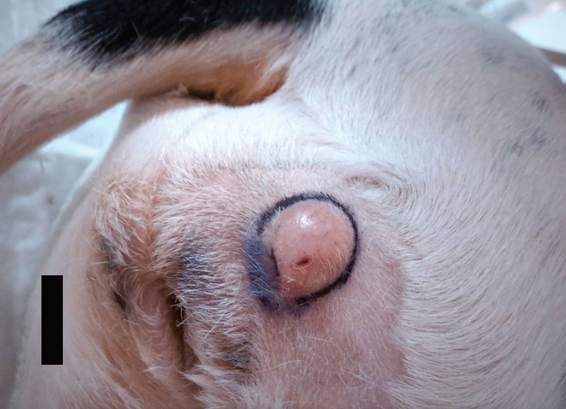
Most dogs with MCTs are clinically well, aside from solitary, or occasionally multiple, cutaneous masses. Masses are most commonly located on the trunk and perineal area (50 per cent), limbs (40 per cent), and head and neck (10 per cent). Mast cell tumours can vary widely in appearance and may be mistaken for non-neoplastic lesions; subcutaneous MCTs can be soft and are often incorrectly diagnosed as lipomas. Low-grade MCTs are commonly slow growing over a period of many months, whereas high-grade MCTs can grow rapidly to a large size and are often ulcerated (see Figures 1 and 2). Clinical signs secondary to mast cell degranulation may be observed including fluctuations in tumour size and peritumoral oedema/erythema, or systemic signs including gastrointestinal upset or rarely collapse. Gastrointestinal ulceration due to histamine release has been reported in 35–83 per cent of dogs with MCTs undergoing post-mortem. Dogs with visceral metastasis can present systemically unwell with neoplastic cavity effusions.

Figure 1. Low-grade cutaneous mast cell tumour lateral to the vulva in a dog.
Prognostic Factors
It is important to understand the prognostic factors associated with canine MCTs prior to further diagnostics and treatment as these factors will likely influence your approach to the case. There are many prognostic factors, and they should be all be considered when assessing a case, as no single factor is entirely predictive of biologic behaviour and outcome.
Histologic grade is the single most reliable prognostic factor when assessing canine cutaneous MCTs. Cutaneous MCTs are routinely graded according to the Patnaik system, as grade 1, 2 or 3, or the Kiupel system, in which tumours are categorised as low or high grade, depending on their histological features. The histological grade of cutaneous MCTs is prognostic; it is well documented that dogs with Patnaik grade 3 tumours have a significantly shorter overall survival time compared with those with Patnaik grade 1 and 2 tumours.9,11 Similarly, dogs with Kiupel high-grade tumours have significantly shorter overall survival times and progression-free survival times compared with those with low-grade tumours. As some tumours within the Patnaik intermediate-grade designation can occasionally behave in a more aggressive manner than expected, the Kiupel grading system is often used concurrently; the high or low Kiupel grade may help predict the biologic behaviour of Patnaik intermediate-grade MCTs. Despite this, some low-grade Kiupel/intermediate-grade Patnaik MCTs can still behave aggressively. For these intermediate-grade tumours, markers of proliferation such as mitotic index (MI, number of mitoses per 10 high-powered field), Ki67, and argyrophilic nucleolar organising region (AgNOR) can help determine if a biologically aggressive behaviour is to be expected. Mast cell tumours with a Ki67 index >23 positive cells per grid have a significantly increased risk of local recurrence, metastasis, and MCT-related death. Similar findings were observed in MCTs with increased AgNOR counts. An AgNOR x Ki67 index >54 has been associated with increased risk of local recurrence and MCT-related death. Mitotic index is a strong independent prognostic factor; one study demonstrated dogs with a MI <5 had prolonged survival (median survival time [MST] 80 months) compared to dogs with a MI >5 (MST three months).
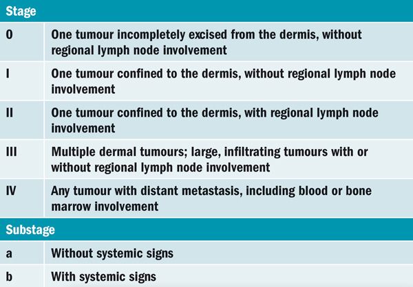
Clinical stage is also predictive for survival; the World Health Organization clinical staging system for MCTs is presented in Table 1. Metastasis is often to local lymph nodes (LNs), liver, spleen and, rarely, bone marrow and lungs. Occasional reports of metastasis to other intrathoracic tissues such as heart, pericardium and mediastinum exist. Increasing stage of disease is correlated with a worse prognosis, with a median survival time of just 110 days in dogs with distant metastatic (stage IV) disease. Metastatic rate increases with tumour grade, with 55 to 96 per cent of dogs with high-grade MCTs demonstrating evidence of metastasis.

Table 1. World Health Organization staging system for canine cutaneous mast cell tumours.
Other prognostic factors include c-Kit mutation status, KIT expression pattern, tumour location, and signalment, although this is not an exhaustive list.
Activating mutations in exons 8, 9 and 11 of the c-Kit gene are common in canine MCTs, with one study demonstrating mutations in approximately 25 per cent of tumours. C-Kit mutations are also significantly associated with higher grade MCTs and are associated with decreased disease-free interval, increased risk of tumour recurrence and decreased overall survival time. The KIT expression pattern is also associated with prognosis. Normally, KIT is expressed in the cell membrane of mast cells, which is classified as KIT staining pattern 1. In some neoplastic mast cells KIT is expressed focally (staining pattern 2) or diffusely (staining pattern 3) throughout the cytoplasm. Increased cytoplasmic KIT expression is associated with increased risk of local recurrence and reduced overall survival.
Tumours located in subungual or perineal/inguinal regions, especially if located on the prepuce or scrotum, have been associated with more aggressive behaviour and should be approached with this in mind. Mast cell tumours of the muzzle are associated with a higher rate of regional LN metastasis (<60 per cent) although long term outcomes can still be favourable in these cases.
Although some breeds of dog including pugs, boxers, and dogs of bulldog descent are predisposed to MCT development they generally seem to develop lower-grade tumours.

Figure 2. High-grade cutaneous mast cell tumour on the digit of a dog, with marked peritumoral oedema and ulceration.
Further Investigations
MCTs are diagnosed easily via fine needle aspirate cytology in most cases. The round cells contain characteristic metachromatic cytoplasmic granules (Figure 3) although sometimes these may not stain with routine Romanowsky-type stains, requiring assessment with either Wright-Giemsa or toluidine blue stain. High-grade undifferentiated MCTs may lack granules and can therefore be challenging to differentiate from other round cell tumours.

Figure 3. Cytology from a cutaneous mast cell tumour showing granulated, well-differentiated mast cells (arrow).
Full pre-operative staging should include haematology, biochemistry, urinalysis, regional LN aspirates and abdominal ultrasound for liver and spleen aspirates (these should be obtained regardless of ultrasonographic appearance as the liver and spleen can appear normal despite harbouring metastatic disease). However, if the mass is in a location amenable to wide surgical excision and lacks any of the aforementioned negative prognostic factors, then surgical excision could be performed initially, with staging undertaken if histology identifies an intermediate-high grade or highly proliferative MCT. Pre-operative cytology or histology via incisional biopsy could be considered to help guide decision making; a cytologic grading system has been devised but it should be noted that both cytologic grading and grading based on pre-operative biopsies can underestimate the true grade of the MCT in a small proportion of cases. Thoracic imaging is rarely indicated as pulmonary metastasis is very uncommon; however, it may be prudent prior to invasive or expensive procedures to rule out other comorbidities. To reduce the risk of MCT degranulation, chlorphenamine should be administered prior to aspiration of the primary tumour, LNs, liver or spleen.
Assessment for LN metastasis should not be based solely on LN size or location. A sentinel lymph node (SLN) is defined as the first LN within a lymphatic basin that drains the primary tumour. Historically, the SLN was often considered the closest LN to the tumour but this is not always the case, especially in oral/head tumours where lymphatic drainage patterns can be unpredictable; one study demonstrated metastasis to contralateral LNs in 62 per cent of cases with malignancies of the head. Another study demonstrated that 42 per cent of dogs with MCTs had a SLN different to the anatomically closest LN. Regarding LN size, one study evaluated extirpation of non-palpable or normal-sized regional LNs in dogs with cutaneous MCTs and found that 49.5 per cent of these LNs were metastatic. Therefore, aspirates of LNs should always be obtained regardless of their size. If there is any doubt over the location of regional LNs, then SLN mapping procedures such as indirect CT lymphography may be performed, although these techniques may not be possible in all general practice settings. Detection of LN metastasis via cytology is between 63 to 100 per cent sensitive and 83 to 96 per cent specific depending on the tumour type, so ideally, LN extirpation for histopathology should be performed, even for LNs with a negative cytology result, as occasionally metastasis can be missed based on cytology alone.
Treatment
The initial treatment of choice for MCTs, where possible, is surgical excision. Historically 3cm lateral margins were recommended for all MCTs and, although this is still advised for high-grade MCTs, 1cm and 2cm lateral margins are acceptable for low- and intermediate-grade MCTs respectively. Regarding deep margins, excision should always include removal of one uninvolved fascial plane irrespective of histologic grade.
If complete surgical excision is achieved for a low- or intermediate-grade MCT, then no further treatment is required. However, routine follow-up is prudent with physical examination to include lymph node assessment advised every three months for the first 18 months, then every six months thereafter. If excision is incomplete, then several options are available. If location allows, re-excision with wide margins is advisable to remove residual microscopic disease. If revision surgery is not possible, then alternative options include adjuvant radiation therapy or chemotherapy. Active surveillance is also an option if adjuvant therapy is not possible, as it is possible the MCT will not recur; only between 10-30 per cent of incompletely excised low or intermediate grade MCTs recur.
Due to their high metastatic risk, adjuvant systemic therapy (chemotherapy or targeted therapies [tyrosine kinase inhibitors]) is always recommended in dogs with high-grade MCTs even where complete surgical excision is achieved. If the mass is incompletely excised, then further local treatment (revision surgery or RT) is also strongly advised due to the high risk of local recurrence compared to lower grade MCTs; if these options are not possible then the systemic therapies employed to reduce the risk of metastatic disease will also target residual local disease. After completion of adjuvant systemic +/- local treatment routine follow-up as previously outlined is recommended. Systemic therapy should also be considered in cases with negative prognostic factors other than just tumour grade, such as high-risk tumour locations or high proliferation indices.
Sentinel/regional lymph nodes should be surgically excised if identified as metastatic during initial staging; as discussed above, LNs should also ideally be removed, even if cytologically negative for metastasis, due to the risk of false negative results. Lymph node extirpation is especially important for high-grade MCTs due to the increased risk of metastasis. Removal of regional LNs has a positive impact on survival in dogs with stage II MCTs, with median survival times in the region of >6 years achievable (compared to around one year in dogs where metastatic lymph nodes were not removed); radiation therapy of regional LNs can also be considered.
If a MCT cannot be excised then local treatment options in the gross disease setting include RT or systemic chemotherapy/targeted therapy. Additionally, tigilanol tiglate (Stelfonta) is a recently approved novel treatment for non-resectable canine MCTs; after intratumoral administration a complete response rate of 75 per cent is reported with 89 per cent of dogs recurrence-free 12 months after treatment. It should be noted that tigilanol tiglate is only licensed for non-resectable, non-metastatic subcutaneous mast cell tumours located at, or distal to, the elbow or the hock, and non-resectable, non-metastatic cutaneous mast cell tumours. Regardless of treatment approach, in any dog where gross disease remains it is important to reduce the risk and consequences of MCT degranulation; the author uses a combination of H1 receptor antagonist chlorphenamine and proton-pump inhibitor omeprazole ongoing for as long as gross disease is present.
There are a variety of chemotherapy options for canine MCT mainly revolving around vinblastine, lomustine, tyrosine kinase inhibitors (TKIs) or combinations thereof. A 12-week vinblastine protocol (administered weekly for four doses, then every other week for four doses) is a well-tolerated protocol for low-intermediate grade MCTs where chemotherapy is indicated (e.g., incomplete excision/microscopic disease setting, high-risk tumour location); a combined vinblastine/lomustine protocol can be considered for high-grade MCTs. In situations where the primary tumour cannot be excised, or treated with RT or tigilanol tiglate, then TKIs such as toceranib or masitinib can be considered with overall response rates between 43 to 82 per cent reported. Good responses to TKIs can be seen in MCTs even if they do not harbour c-Kit mutations, although response rates are often higher in cases that do. Prednisolone inhibits mast cell proliferation, incudes cell apoptosis and reduces peritumoral inflammation and oedema so is used alongside chemotherapy in the treatment of MCTs. It can also be used alone in a palliative setting if required, with an overall response rate of 20 per cent reported.
Chemotherapy is usually well tolerated and if adverse effects are seen they are often mild, including gastrointestinal toxicity and myelosuppression. In addition, lomustine can cause cumulative hepatotoxicity and TKIs can induce hepatotoxicity, hypertension and proteinuria, all of which need to be closely monitored for throughout treatment. This list of chemotherapy protocols is by no means exhaustive and, as treatment recommendations depend highly on histologic prognostic factors, disease stage, and independent patient factors, consultation with a veterinary oncologist is sensible before starting chemotherapy. Patient-specific monitoring can then also be advised depending on treatment protocol.
Conclusion
Although most MCTs encountered in general practice are low-grade and can be cured with complete surgical excision, more aggressive and complex cases can be seen. A thorough understanding of prognostic factors for canine MCTs is therefore important to help identify these cases and plan staging and treatment accordingly. The presence of regional metastatic disease should not preclude further treatment as good outcomes can still be achieved in many of these cases. For non-surgical MCTs, there are still a variety of treatment options available including radiotherapy, chemotherapy or tigilanol tiglate.
References available on request.
1) Approximately what percentage of canine mast cell tumours will harbour c-Kit mutations?
A. 15 per cent
b. 25 per cent
c. 35 per cent
d. 45 per cent
2) What are the minimum lateral surgical margins recommended for a low-grade cutaneous mast cell tumour?
A. 0.5cm
b. 1.0cm
c. 1.5cm
d. 2.0cm
3) Hypertension is a potential adverse effect of which chemotherapy agent?
A. Lomustine
b. Vinblastine
c. Toceranib
d. Tigilanol tiglate
4) Approximately what percentage of canine mast cell tumours will respond (reduce in size) to treatment with prednisolone alone?
A. 20 per cent
b. 30 per cent
c. 40 per cent
d. 50 per cent
5) Mast cell tumours in which location have an increased risk of lymph node metastasis?
A. Distal limb
b. Lateral thorax
c. Subcutaneous
d. Muzzle
Answers: 1B; 2B; 3C; 4A; 5D.


