Open fracture management in small animals
European specialist in small animal surgery Fernando S. Reina Rodriguez DVM, MSc,DVMS, DipECVS and small animal surgeon Jose V. Targa Villalba DVM, Cert. VEAMIS, Cert. Orthop.UCH-CEU offer some guidelines on open fracture management
Open fractures occur when a fracture and an open wound happen simultaneously or there is a loss of integrity of the skin near the broken bone. According to some authors, the incidence of open fractures in small animals is low; however, others have reported a much higher incidence, which can reach up to 5–10 per cent of all fractures.
Contrary to other orthopaedic disorders, open fractures are considered surgical emergencies. This article describes basic guidelines for their classification, initial management, and treatment to improve the outcome and maximise the chances of returning to normal function.
Introduction
An open fracture involves bone that is exposed to environmental contamination because of loss of soft tissue integrity. These fractures are generally caused by high-energy trauma, and often the wound occurs by a combination of direct trauma to the soft tissues and fragments of bone breaking through the skin at the time of injury. The most commonly affected anatomical locations are those with less soft tissue coverage, such as the distal part of the extremities (Figures 1 and 2). Due to the traumatic nature of these fractures and the high incidence of complications such as osteomyelitis or failure of fracture repair, open fractures are considered surgical emergencies.
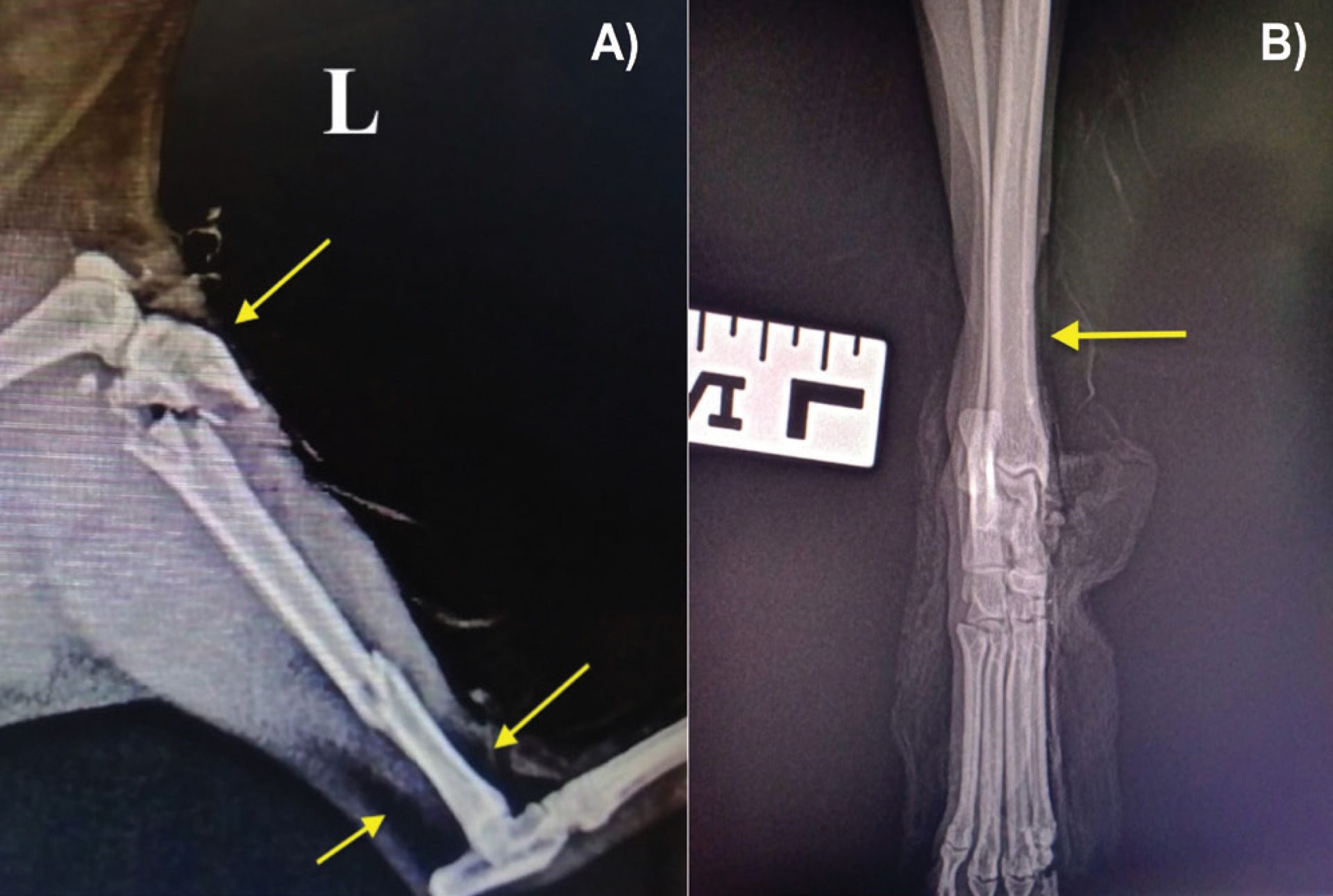
Figure 1: Examples of radiographic appearance of open fractures. A) Highly comminuted fracture in a feline patient affecting the proximal and distal diaphysis of the left tibia following a fall from a third floor. B) Left tibiotarsal mediolateral subluxation (view in neutral position) in a canine patient after a road traffic accident. The yellow arrows indicate gas accumulation under the skin due to soft tissue barrier disruption.

Figure 2: Examples of radiographic appearance of open fractures. A) Caudo-craneal and B) mediolateral radiographs of a comminuted fracture of the mid diaphysis of the right tibia in a five month-old canine patient after a road traffic accident. The yellow arrows indicate gas accumulation under the skin due to soft tissue barrier disruption.
Initial patient management
Primary treatment is focused on recognising and treating injuries other than the fracture that could compromise patient survival. Open fractures are caused by high-energy trauma such as traffic accidents or falls from a high location; for this reason, a general physical examination with a focus on evaluating ABC (airway, breathing and circulation) must be a priority. The wound can be covered with a sterile dressing until later evaluation is performed. Once the patient is stable, has received adequate emergency treatment (IVFT, analgesia, or oxygen therapy if required) and no life-threatening conditions are identified, initial management of the open fracture can be established.
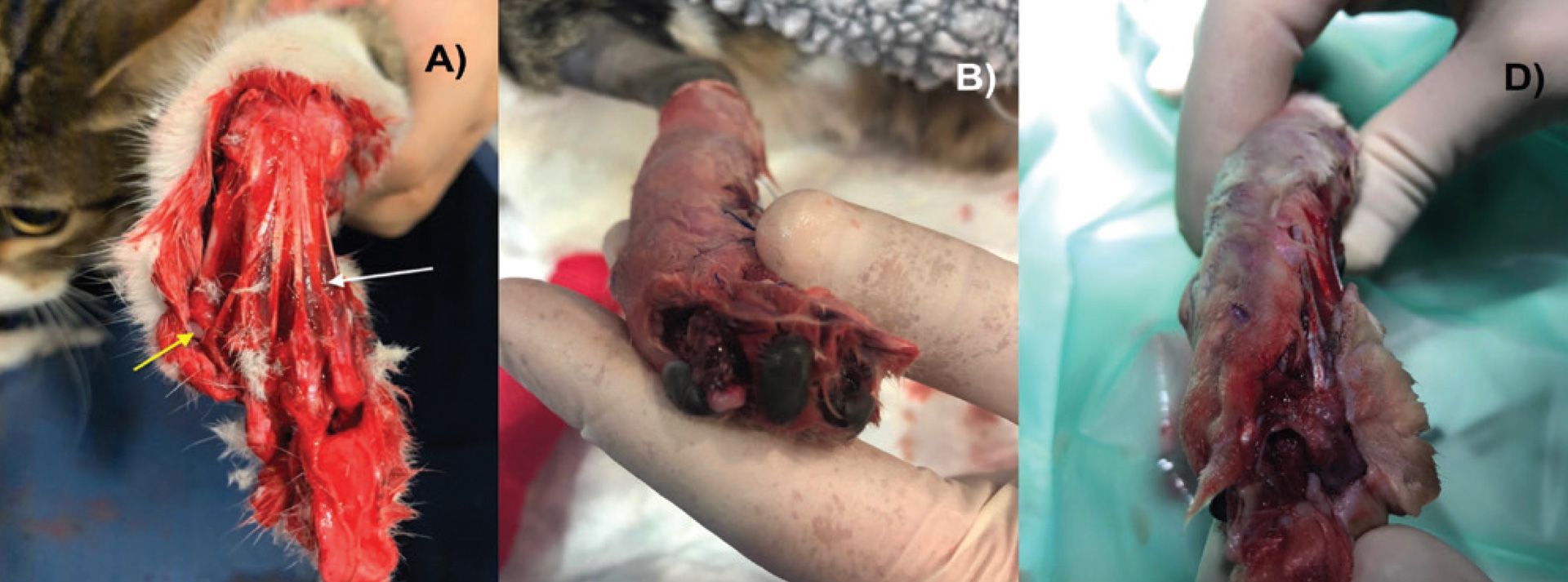
Figure 3: A) Type III open fracture luxation at the metacarpophalangeal joint (yellow arrow) of the left forelimb in a feline patient. Observe the inadequate clipping and small areas of blood congestion (white arrow). B) and C) 24 hours after presentation, the limb became markedly cyanotic and cold. A forequarter amputation was performed.
It is important to perform a neurological examination before wound assessment, as the presence of neurological deficits will affect decision-making. Thereafter, the extension of the wound and fracture can be evaluated. Wound assessment should be done under general anaesthesia or deep sedation. Inadequate clipping (Figure 3A) is a common mistake that causes improper evaluation of the injury and viability of the soft tissues, and it contributes to infection via colonisation of the wound by resident microbes and the presence of organic debris. Applying a small amount of sterile gel over the wound helps avoid further skin damage during clipping.
During wound assessment, it is important to check for adequate blood perfusion, as it is critical for wound healing and outcome. The absence of an arterial pulse, presence of cold temperature, and other macroscopic changes such as congestion and cyanosis (Figures 3B and 3C) may indicate loss of tissue viability. Sometimes it may be better to reassess the area 12–24 hours later, as further changes may occur. Doppler ultrasound has been used in humans to provide an objective evaluation of the extent of vascular damage. It has also been used in small animals to assess bone healing.
Open fracture classification
All systems to classify open fractures are based on the extent of tissue damage. The classification aims to provide an objective evaluation of the wound, avoiding inter-observer variation and establishing the best treatment recommendations. However, fracture classification cannot predict the outcome, as patient factors that influence fracture and wound healing are not considered. The most commonly used is the Gustilo–Anderson classification, which is simple, easy to use, and able to give an idea of the expected outcome and guide treatment regimes (Table 1).
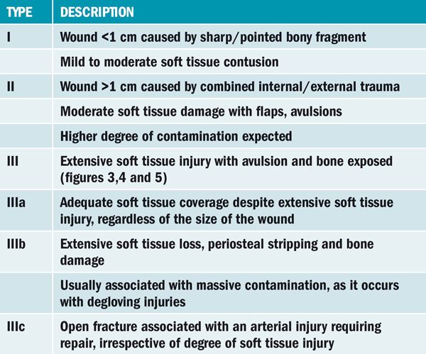
Table 1. Gustilo–Anderson classification for open fractures. Type IIIc fractures are less common in small animals due to extensive collateral blood supply; however, they may occur if initial wound management is inadequate.
Wound management
Adequate early wound management is required to improve outcomes. As previously mentioned, it must be performed under anaesthesia or deep sedation. Manipulation of the area should be done under aseptic conditions to prevent nosocomial infection. This generally requires the use of sterile gloves, drapes and surgical instruments, particularly if surgical debridement is needed.
The use of disinfectants such as chlorhexidine or povidone may be beneficial on the peri-wound skin; however, there are no benefits to using them directly on damaged tissues as they cause irritation when not used at adequate concentrations. Irrigation with standard saline solution provides similar results and fewer side effects. An important factor to consider is the pressure exerted over the wound and open fracture during irrigation, as high pressure may cause further tissue damage and promote bacteria penetration into the deeper tissues. If the exact irrigation pressure cannot be measured, using a 1L plastic bag of fluids within a cuff pressurised to 300mm Hg (15) or a 19G needle attached to a 20mL syringe are cheap methods to ensure adequate pressure is exerted during irrigation.
Debridement of nonviable tissue is the most important factor in preventing infection and ensuring the wound does not remain in an inflammatory phase (Figures 4 and 5). The goal is removing devitalised tissue, which serves as culture for bacterial growth, and improving blood perfusion, oxygen tension, and number of inflammatory cells in the wound. Surgical debridement of skin, subcutaneous tissue or muscular fascia can be performed in a more aggressive manner. Macroscopic changes, such as dark discolouration, cut edges that do not bleed, leathery texture or maceration, are clear signs of nonviable tissue. Debridement of muscle and tendons requires a close examination following the rule of the ‘four Cs’: colour, consistency, circulation and contractibility. Fragments of small tendons should be removed, while larger tendons are best preserved if possible. Bone fragments attached to muscle should be carefully manipulated; however, those completely loose should be removed to avoid development of a sequestrum. When large areas of cortical bone are exposed without any soft tissue coverage, osteostixis may be performed, which involves drilling small holes into the exposed bone to promote granulation tissue formation. Following surgical debridement, further wound lavage should be done to remove detritus and blood clots.

Figure 4: A) Degloving injury with metacarpal fractures (yellow arrows) of the right forelimb in a Greyhound. After initial wound debridement (B), the fractures were repaired with IM pins and the wound managed with daily dressing changes until soft tissue covered the exposed bone (C and D)

Figure 5: A) Type III open fracture/luxation after a road traffic accident. The patient presented with extensive soft tissue loss at the medial aspect of the limb from the stifle (white arrow) to the tarsocrural joint (yellow arrow). Wound appearance 24 hours after surgical debridement at the level of the stfile (B) and tarsocrural joint (C).
Antibiotic therapy
Early antibiotic administration is recommended for all open fractures as it improves outcomes. Administration of antibiotics within three hours post-trauma significantly reduces the incidence of infection compared to cases receiving later antibiotic therapy. Although there is no consensus regarding which drug or drug combination is ideal, the clinician must consider a broad-spectrum antibiotic, effective against both Gram-positive and negative bacteria, and that reaches adequate concentrations at the bone and wound beds. This is even more important for type III fractures, where extensive tissue damage increases the risk of contamination. First-generation cephalosporins administered intravenously are a good therapeutic choice.
It is uncertain what the optimal length of antibiotic therapy is; however, a minimum of 72-hour coverage followed by an additional three days if needed is recommended. In our experience, some cases may require prolonged antibiotic therapy until radiographic healing of the fracture occurs.
If fracture repair and wound closure are not delayed, it is recommended to obtain tissue samples and submit them for culture and sensitivity. However, if the defect is closed by second intention healing or fracture repair is delayed, bacterial cultures may not be crucial, as the initial isolated microbes may differ from those found later.
Fracture fixation
Although there are different methods for treating open fractures, a general rule is to choose a repair that not only provides adequate fracture stability but also allows wound assessment and preserves the integrity of the remaining viable tissues. Factors such as the type of fracture, anatomical location, surgical approach required and vascularity of the skin must be considered before choosing a method of fixation. Because fracture instability is a mechanical factor contributing to osteomyelitis, it is important to choose a method of repair that provides maximum stability. Bone fractures may heal in a contaminated environment as long as there is adequate interfragmentary compression and frame stability. Unstable methods lead to interfragmentary micromotion when loads are applied, contributing to soft tissue necrosis, osteomyelitis, delayed unions or non-unions.
Linear or circular external fixators are common methods chosen to repair open fractures. Compared to internal fixation, this method allows a more biological approach by preserving the local soft tissues and avoiding vascular compromise, which is particularly important when treating open fractures. They allow easy access to the wound bed to evaluate soft tissue healing and make changes in open wound management if needed. They also stand out for their versatility, as they can be successfully used to temporarily immobilise a joint if necessary (Figure 6). They can also be removed once the fracture healing has completed, without any need for a second surgical procedure. Despite the clear advantages of external fixation, some fractures, such as highly comminuted or articular fractures, may require stronger methods of internal fixation (Figure 7). In these cases, a second surgical procedure to remove implants may be required once fracture healing occurs to prevent implant-associated infections.
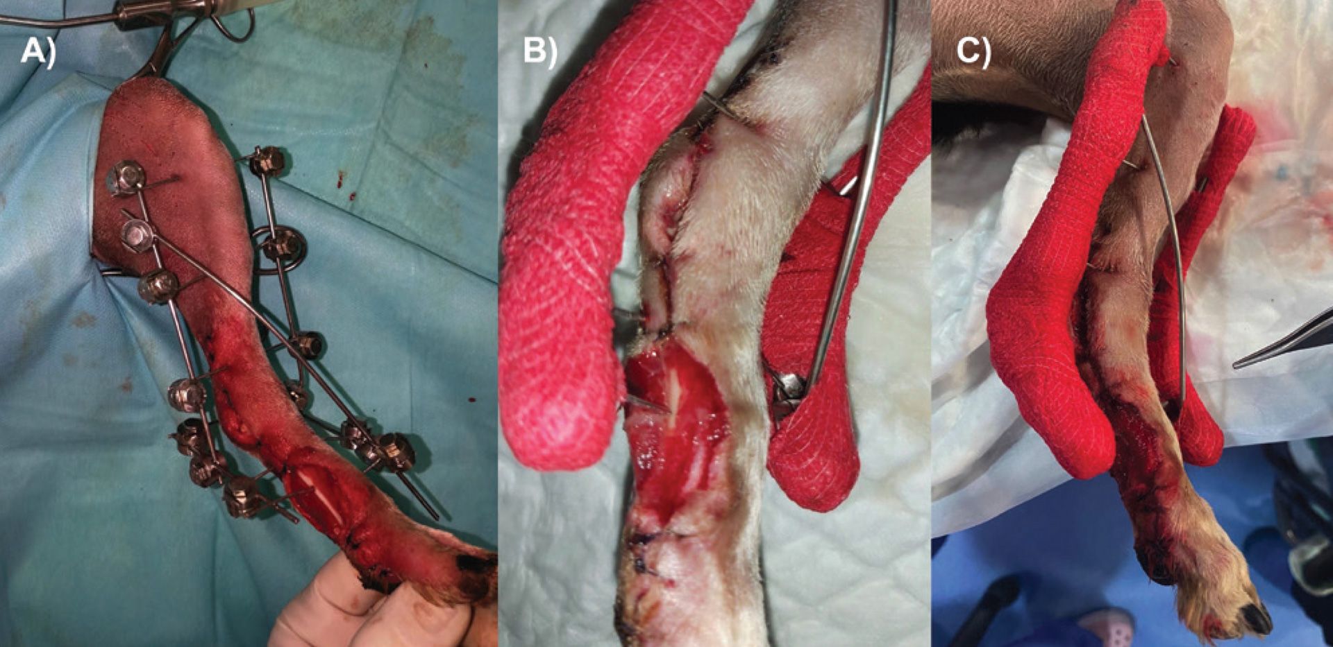
Figure 6: A) Transarticular external fixator placed in the right pelvic limb of a canine patient. This system allows temporary joint immobilisation and easy access to the wound for assessment and management B) and C).
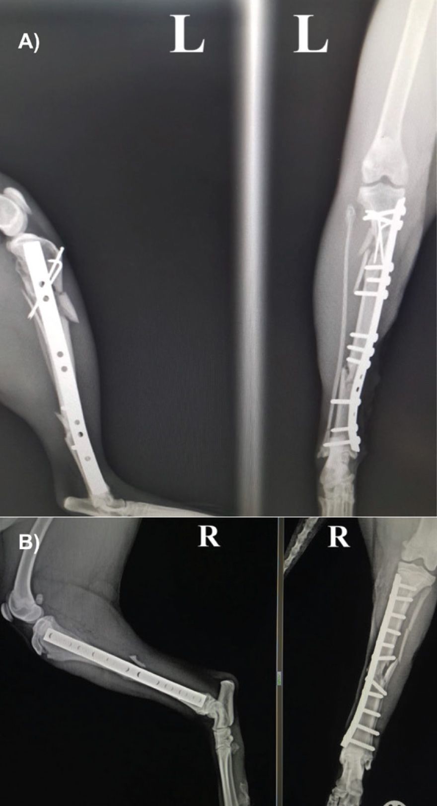
Figure 7: A) Fracture repair in patient of Figure 1A using internal fixation. B) Fracture repair of an open fracture using internal fixation in another feline patient.
Independently of the system selected, the keys for a successful outcome are:
1) choose a method that provides adequate stability to the fracture,
2) achieve anatomical alignment, and
3) minimise soft tissue damage to allow early return to limb function.
Although external coaptation can be initially used to avoid movement of the fracture fragments on presentation, this method should not be used for primary fracture repair.
To minimise the incidence of delayed or non-unions, we can consider the use of bone grafting. Grafts can be placed into a contaminated or infected site; however, bacteria may affect graft survival, so using them in a second procedure may be considered. Although a cortical graft can be used to fill a large gap at the fracture site, it may contribute to development of a sequestrum. For these reasons, it is recommended to delay bone grafting until the wound and fracture sites have improved, even if this approach requires a second surgical procedure.
Outcome and complications
Limited information is available regarding the outcomes and complication rates for open fractures in small animals. In humans, complications such as persistent infection, osteomyelitis or development of delayed/non-unions seem to be directly related to the extent of the injury and are significantly higher for type III fractures. Although it is considered a salvage procedure, limb amputation may be necessary if the extent of the trauma is so severe that there is little or no chance for fracture repair or in fractures with severe postoperative complications. Owners must be informed about this possibility, as some patients require more than one surgical procedure.
If proper management has been done in the early period following the traumatic injury, the outcome for open fractures is generally good. However, many patients require numerous postoperative rechecks to assess wound and fracture healing, limb function and to evaluate the presence of residual infection.
Conclusions
- Open fractures are serious injuries, and they must be considered orthopaedic emergencies.
- A successful outcome depends on adequate initial management.
- Early antibiotic therapy, with or without bacterial culture, is always indicated.
- Initial management includes adequate wound assessment, surgical debridement of nonviable tissues, lavage with isotonic solutions and fracture immobilisation.
- Fractures must be repaired with simple methods that do not disrupt the remaining soft tissues and provide adequate fracture stabilisation.
- Considering the higher incidence of postoperative complications compared to closed fractures, owner communication is crucial as some patients may require a second surgical procedure or even limb amputation when trauma is severe.
References are available on request.
1) Which of the following statements is true?
a. Only type III open fractures are considered surgical emergencies due to the extensive soft tissue damage
b. Only the Gustilo-Anderson classification takes into account patient-dependent factors that affect wound and fracture healing
c. The use of cephalosporins is a good choice to treat open fractures if culture and sensitivity tests are not available
d. All the above are true
2) Regarding the use of external fixators to treat open fractures, which of the following statements is NOT true?
a. They require a minimal surgical approach, respecting the remaining soft tissues
b. This method is recommended for articular fractures and highly comminuted fractures where more rigid fixation is required
c. They can be used to temporarily immobilise a joint
d. They allow direct wound assessment and management even after fracture repair has been done
3) Which of the following is true in relation to the use of antibiotics for open fractures:
a. The use of antibiotics is always recommended. They need to be administered for a minimum of 72 hours at the beginning followed by an additional three days if required
b. The use of antibiotics improves the final outcome, particularly if they are administered within three hours since the trauma takes place
c. In the absence of culture and sensitivity results, it is better to select an antibiotic effective against both Gram-positive and negative that reaches adequate concentration at the wound and fracture beds.
d. All of the above are true
4) Which of the following are true regarding wound management for open fractures:
a. Chlorhexidine or iodine solutions are better for cleaning and flushing the wound bed due to their disinfectant properties
b. Diluted antibiotics are better for cleaning and flushing the wound bed due to their antibacterial properties
c. Irrigation with standard saline solution, applying the adequate pressure during lavage, provides similar results and fewer side effects
d. All of the above are correct
5) Which of the following statements is true regarding the postoperative care of open fracture fixation?
a. Some patients may require prolonged rechecks and follow up radiographs
b. It is important to monitor for potential complications such as osteomyelitis, delayed unions or non-unions.
c. The rate of postoperative complications is significantly higher compared to close fractures
d. All of the above are true
Answers: 1C; 2B; 3D; 4C; 5D.


