Dental abscess in a chimpanzee: a case study
John Breen BSc VN, regional sales manager with iM3 Dental, reports on the treatment of a dental abscess in a chimpanzee from a troop in Dublin Zoo
Marlon, a 26-year-old chimpanzee in Dublin Zoo, is the alpha male in a troop of five females and two males. Zookeepers raised concern when Marlon became standoffish with his peers. He distanced himself from group activities, ate alone and challenged troop members when approached. The hierarchy system among chimpanzees is complex and if an alpha male displays signs of weakness, another male can seize this opportunity to assert his power for the title. It was clear the issue required early attention for Marlon’s welfare and the troop’s politics.
Not being able to conduct a thorough conscious clinical examination makes diagnosing zoo animals difficult. Often, the only information at the vet’s disposal is the animal’s behaviour and observable symptoms. With that in mind, zookeepers closely observed Marlon to discern any gross evidence of pathology and subsequently, they noticed a swelling beneath his right eye which would be consistent with a tooth root abscess. At this point, Dr Emma Flynn, one of Dublin Zoo’s resident vets, and her team knew they had reason to intervene and began to make suitable arrangements to treat Marlon.
Preparation
Organising any surgical procedure for a zoo patient requires coordination and planning. Anaesthesia was provided by University College Dublin who have worked closely with Dublin Zoo for all their medical procedures. Dental radiographs and equipment were provided by iM3 Dental who specialise in veterinary dentistry equipment. Dr Nora Schwitzer, the veterinary surgeon who led the procedure, was joined by human dentist, Dr Thomas Linehan of Seapoint Dental Clinic. As an evolutionary cousin of humans, a chimp’s dentition has a closer resemblance to our own teeth than to those of cats or dogs, so Dr Linehan’s insight helped identify techniques and explain observations not normally used or seen in general veterinary practice.
Marlon weighed 75kg and it was clear that transporting him to surgery was not going to be easy. Safety and Marlon’s welfare were top priorities. It was also important to everyone that Marlon wake up in familiar surroundings during his recovery to make it as stress-free as possible for him. Since the chimpanzee enclosure is a fair distance from the zoo’s in-house surgery and the pathways between the two are filled with visitors during the day, it was decided to create a pop-up surgery room in the chimpanzee enclosure where Marlon could be treated and then be monitored in recovery by the zookeepers who know him best.
Anaesthetic Protocol
Kyratsoula Pentsou and Joanna Potter from UCD were the head anaesthesiologists for Marlon’s operation. Oral midazolam was administered to the patient prior to the procedure to provide anterograde amnesia. Following this, immobilisation was achieved with intramuscular administration of medetomidine and ketamine via a dart. Once intravascular access was obtained and the patient’s trachea was intubated with an endotracheal tube, anaesthesia was maintained with intravenous infusions of propofol, fentanyl and ketamine inhalant combined with isoflurane administered in 100 per cent oxygen. A multiparameter monitor was connected to the patient and was used to continuously monitor and assess oxygen haemoglobin saturation, end-tidal carbon dioxide levels, arterial blood pressure values, temperature, electrocardiographic and capnographic changes, respiratory rate, heart rate and inhalant agent levels. The patient was breathing spontaneously throughout the procedure while the only complication noted was mild hypotension which was treated successfully with the administration of lactated ringer’s solution, a colloid bolus, dobutamine infusion and intravenous ephedrine. A mandibular block, a palatine block and an infraorbital block with bupivacaine 0.5 per cent were performed to optimise patient comfort and intravenous paracetamol was also added to the protocol for the same purpose. Prior to recovery, intravenous maropitant was administered to decrease post-operative nausea. Intramuscular atipamezole was used to antagonise the medetomidine sedative effects once the patient was placed securely in the recovery area. Recovery was uneventful and the animal resumed normal function the same afternoon.
Diagnosis
Once Marlon was induced and stable, Dr Schwitzer began her oral examination. She was able to immediately confirm that the presenting problem was a periapical tooth root abscess that had since burst with visible pus discharge from the mucogingival junction (See Figure 1). The abscess stemmed from tooth 104, the right maxillary canine which had suffered an open fracture, exposing the delicate pulp and resulting in the infection. Further examination of the mouth showed three more complications; a fractured mandibular canine (304), fractured maxillary canine (204) and the early development of a tooth root abscess on one of the incisors (301).
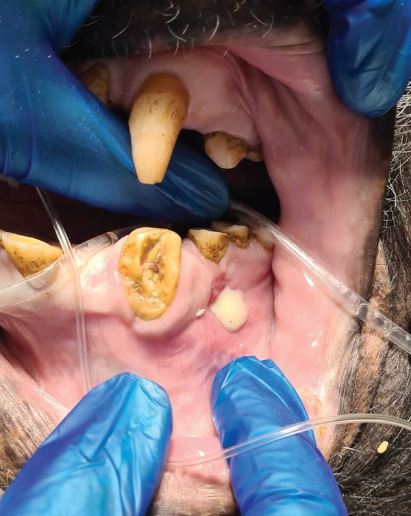
Figure 1. Abscess stemmed from tooth 104 with visible pus discharge from the mucogingival junction.
Procedure
As the first step before beginning any surgical work is to take dental radiographs, a full set of mouth x-rays were carried out (Figure 2). Radiography is a crucial step to get a complete picture of the tooth and assess any pathologies lurking beneath the observable region. Up to two-thirds of a tooth can sit beneath the gumline and any number of hidden pathologies could be present. Large teeth, such as molars and canines, occupy a significant space in their respective jaw segments.
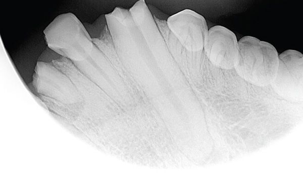
Figure 2. Pre-operative dental x-ray.
Large teeth, such as molars and canines, occupy a significant space in their respective jaw segments. Because of the substantial surface area involved, there is considerable scope for these hidden pathologies to exist. Therefore, careful planning is crucial so that any problems can be noted and considered in the surgical plan.
A bisecting angle technique was used to capture radiographs of each quadrant in Marlon’s mouth. Marlon had to remain in sternal recumbency but achieving the correct angles was possible using iM3’s positioning kit. These angle guides provided both the angle and distance to take the x-ray, ensuring tooth roots were captured and minimising any scatter in the surrounding environment. As the focus was on the canines, a 70-degree angle was used to attain a steep inclination and avoid any elongation that would push the roots off the plate. The radiographs (Figure 3) confirmed complicated fractures in three of the four canine teeth. Factoring in the size of the canines, surgery time, prognosis and Marlon’s welfare, extracting the affected teeth was more practical and therapeutic than endodontic repair.
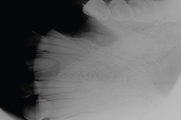
Figure 3. 70 degree bisecting angle technique used to capture full canine root on a single image plate
Dr Schwitzer began with the presenting problem of the 104 canine and created a gingival flap. This is a standard approach when extracting canines in any species as they are large, long teeth and their curvature makes it difficult to fully luxate the periodontal ligament and elevate the tooth from the alveolar bone. Post-extraction, this flap also acts as a bandage that can be sutured back into position to better manage complications like dry socket and speed up the healing process. This is helpful since an open wound could lead to post-op complications. Any need for future repeat surgery was to be avoided due to the difficult logistics of zoo surgeries.
Next, Dr Schwitzer began removing the overlying bone using a surgical length bur and iM3’s LED Advantage High Speed Handpiece. Removing the bone did not come without difficulty as the bone around the tooth was dense which meant the tooth was deeply encased. It was crucial that this bone was adequately removed as attempting to extract a tooth without detaching it from surrounding bone and ligament massively
increases the risk of iatrogenic jaw fractures or fractured teeth.
Precise channels were carved on the rostral and caudal edges of the canine on the buccal side of the tooth. Some bone was also removed from the palatal side which is usually avoided in veterinary as the palate is flat in cats and dogs and it would be easy to accidentally penetrate the nasal cavity. Fortunately, the palate of a chimpanzee is arched like that of a human, so this approach is feasible. After ten minutes of drilling, it became clear that there was a degree of ankylosis. This is often found in teeth that have periodontal disease as bone replaces the regions of necrotic tissue in a response to the infection. After the tooth was carefully detached from the surrounding bone and ligament, the real ‘fun’ began. An elevator was used to painstakingly elevate the tooth from the socket. This was a slow and energy-expending exercise due to the shear length of the chimpanzee’s canine.
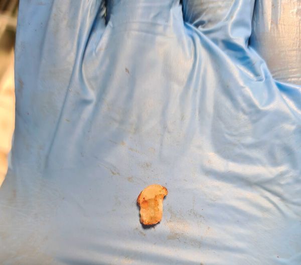
Figure 4. Removed fragment of overlying bone.
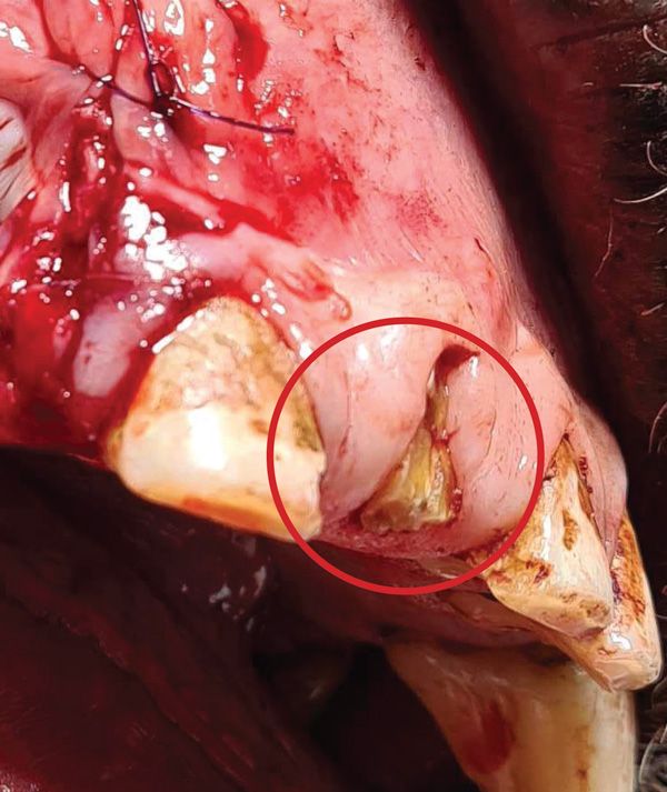
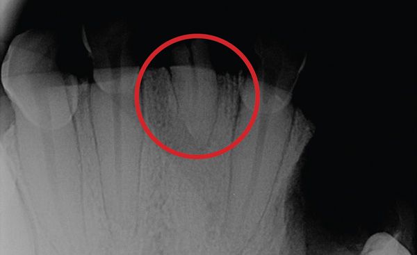
Figures 5 and 6. 301 incisor, unremarkable to the naked eye, but radiographs showed the formation of an abscess.
The extraction of the first canine took approximately 50 minutes, excluding the overall survey of the mouth, dental x-rays, and suturing. It was then time to tackle the 204 and 304 canines. The same approach was used as for the 104 except this time, in the case of 304, there was the added complication of a fractured root.
This was initially hard to confirm by x-ray as the fracture point almost perfectly aligned with the circumference of the jaw line, superimposing it against the bone. It was, however, palpable using a curette inside the socket. Further luxation and elevation meant it could be extracted in one piece. It was easy to confirm this was the full root as there was a ‘halo’ of smooth bone that was consistent with granulation tissue forming around it.
Lastly, the 301 incisor was extracted. At first glance, this tooth was snug and did not protrude significantly. It was unremarkable to the naked eye, but radiographs showed the formation of an abscess. Three hours into surgery, a complication-free extraction of this incisor was very welcome.
Conclusion
Dental disease and pathologies are an inevitable occurrence across all species. Diet plays a significant role, and this is perhaps the largest risk factor in humans. Moreover, lack of prophylactic management predisposes animals to dental conditions and dental trauma can often cause tooth fractures and luxation requiring veterinary intervention.
Marlon made a full recovery and, as he was now much more comfortable, zookeepers could see the improvement in his behaviour immediately. A post-op check was carried out 10 days later by Dr. Schwitzer with very impressive results. Due to the direct blood and nerve supply teeth have, the pain associated with dental disease can be debilitating. Early intervention is always key. The staff at Dublin Zoo were quick to act, and we can be very hopeful that Marlon will continue his reign in the African Plains.
You can catch Marlon’s full story on RTE later this year.
A special thank you to Dr Nora Schwitzer, Dr Emma Flynn, Kyratsoula Pentsou and Joanna Potter for their contributions to this article and to Laurie Harrison of iM3 for editorial help.


