


Dental radiography – giving you the bigger picture
Dental radiography is an essential tool in helping to identify, diagnose and treat oral health issues and underlying dental problems. Here, Ingela Ericsson BA BSc VN, provides an overview of the role of technology in managing and future-proofing oral healthcare
Introduction
Lola is in for her annual booster and, on the detailed oral exam, you diagnose tooth resorption on the feline patient’s left mandibular third premolar, 307. How do you determine the best treatment option? On another oral examination, you diagnose a complicated crown fracture with pulpal exposure and necrosis for the maxillary right fourth premolar, 108, in a canine patient. How do you determine whether there are root fractures, root resorption or periapical disease before planning your treatment? The answer to both those questions is dental radiography.
Oral health
Full mouth dental radiographs should be an essential part of any complete oral health assessment (COHAT). The knowledge gained from dental radiographs not only improves patient care, it increases client compliance with treatment recommendations and, bottom line, will increase your revenue stream from dentistry, improve time management and greatly reduce stress. Dental radiography allows you to deliver high-quality dental care for patients and enables you to:
- Reach a diagnosis;
- Assess extent of pathology;
- Plan optimal treatment;
- Perform certain procedures;
- Assess outcome of treatment performed.
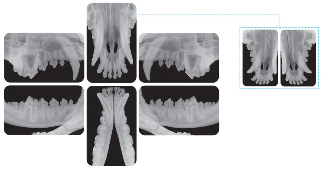
Figure 1: Full mouth dental radiographs should be an essential part of any oral health assessment.
Because of the nature of the anatomy in the oral cavity and the fact that a large proportion of the tooth is hidden below the gumline, coupled with the fact that not all patients are all that willing to allow their mouths to be examined, many disease processes can go unnoticed. And even when there is an obvious issue, there is only so much you can diagnose simply by looking into the patient’s mouth. The visible component of a tooth only comprises approximately 30% of its entire length. The root structure, embedded in the alveolar bone, makes up the rest and the vast majority of periodontal, endodontic and dental pathology is hidden below the gingival margin.
A study looking at the clinical relevance of dental radiography in cats and dogs, where no clinical findings were present on oral examination, showed that, even with visibly normal and healthy teeth, 42% of cats and 28% of dogs had clinically relevant dental disease identified radiographically. More importantly, with clinical indications of dental pathology, additional findings of 50% in dogs and 54% in cats were made using dental radiography (Verstraete et al, 1998).
Table 1: Value of radiographs when no clinical findings are present.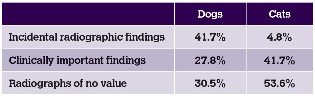
Table 2: Value of radiographs when clinical findings are present.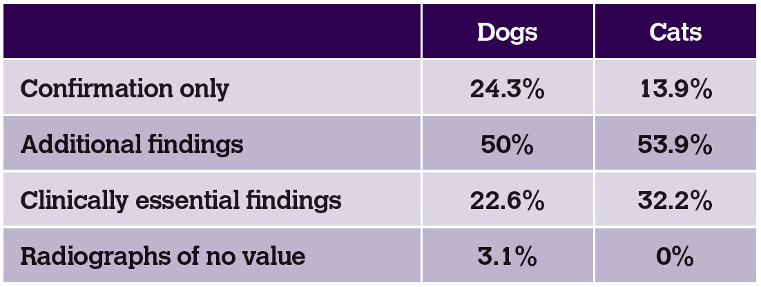
The aim of dental radiography is to produce a diagnostically acceptable full mouth overview as a reference for any subsequent images in the future. The overview enables you to visualise the entire length of every tooth, including the surrounding bony anatomy and gives you a complete picture of the oral status of the patient. A full general anaesthetic is required and it is advised to stage the dentistry work into two, if not three, parts to reduce the risks associated with prolonged anaesthetic times, enable a full and transparent discussion with the owner in terms of treatment needs and costs and allow for a planned and timely procedure.
Plan
The first stage involves the patient being admitted for a full COHAT. This includes dental descale, polish, dental radiography, charting and any emergency work needed (this does not mean you do a full dental on the day). Based on the results, you sit down with the client and clearly and concisely show and explain the findings, help them budget for the work needed and make arrangements for follow-up dental work within four to six weeks. This allows you to better prioritise and time-manage the surgical dentistry work that will follow and greatly reduces stress as you have been able to make an informed decision about treatment needs. It also allows the client to financially prepare for the procedure, making them much more likely to follow recommendations. Finally, it greatly reduces the risks associated with prolonged anaesthetic procedures and improves animal welfare since you will have correctly diagnosed pathology and prioritised your work.
Technology and technique
Correct equipment and technique are essential if your dental radiography work is to be efficient, safe and profitable. The dental radiography generator needs to be manoeuvrable and located close to the dental area. Unlike full body imaging, dental radiography relies on multiple images being taken throughout your dental procedure to allow for evaluation of progress and confirm clinical work. Moving the patient to a different location each time an image is taken is not only inefficient but also has a potential impact on patient safety. Most dental radiographic generators can be either wall mounted close to the workstation or used as a mobile unit.
Your developer or scanner along with a high-quality large computer screen should also be within easy access to your workstation to allow you to use the full mouth overview and any additional images taken as reference during work.
Patient positioning should be consistent, and the radiographs taken in a systematic manner, incorporating every tooth in the maxilla and mandible. This eliminates variables in the quality of work. Different systems to catch radiographic images are available and the two most common types are DR and CR technology. DR technology relies on a small sensor, attached via a cable to the computer. Advantages of DR include that it immediately produces an image on your screen and is relatively cheap to purchase. Disadvantages of the system include the small and bulky size of the DR sensor itself, the difficulty in correct positioning and the need for a large number of exposures in order to gain a full mouth overview.
CR technology, on the other hand, relies on thin photosensitive phosphor-coated plates that are fed into a developer post-exposure. The advantages of CR include the ease with which it can be used and positioned, the ranging plate sizes which helps reduce the number of exposures needed and overall anaesthetic time. The main disadvantage is the wide-ranging products with varying levels of quality available on the market.
The other critical part of dental radiography is learning about positioning techniques and radiographic interpretation. There are a number of books, courses and online resources available to improve your skills and there are also some excellent positioning aids to quickly get you up to speed.
Conclusion
Nearly every patient seen in practice needs or will need regular dental treatment at some stage in their life. Dental radiography provides critical information for both diagnosing and treating dental disease and allows for a planned approach. In addition, dental radiography helps improve animal welfare, increase client compliance, boost revenue, enable better time management and reduce stress.
- Verstraete F, Kass P and Terpak C. 1998a. Diagnostic value of full-mouth radiography in dogs. American journal of veterinary research, 59, 686-91.
- Verstraete F, Kass P and Terpak C. 1998b. Diagnostic value of full-mouth radiography in cats. American journal of veterinary research, 59, 692-5.